Copyright
©2011 Baishideng Publishing Group Co.
World J Gastroenterol. Dec 21, 2011; 17(47): 5227-5230
Published online Dec 21, 2011. doi: 10.3748/wjg.v17.i47.5227
Published online Dec 21, 2011. doi: 10.3748/wjg.v17.i47.5227
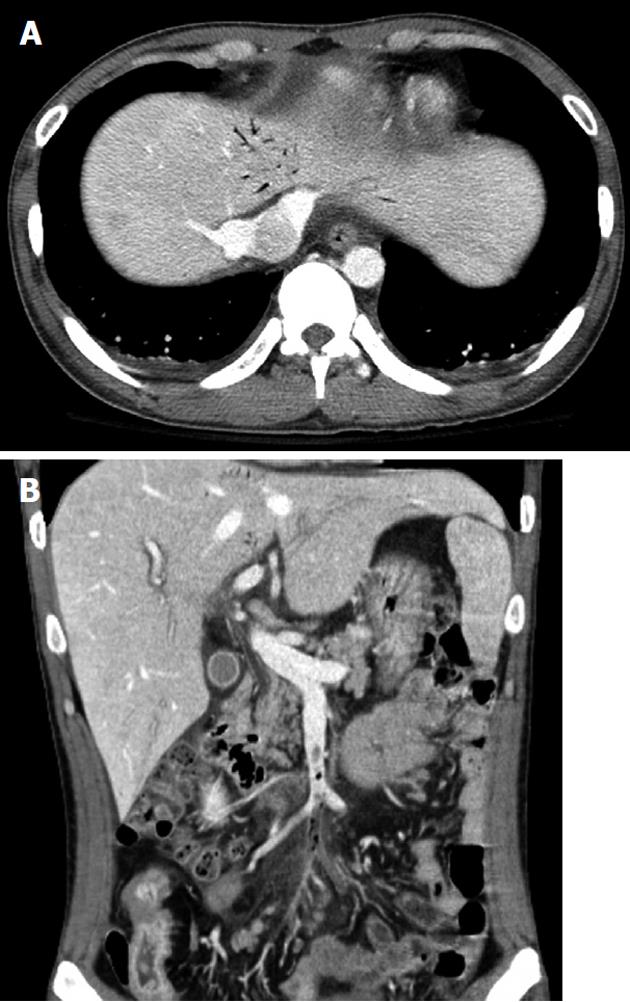
Figure 1 Computed tomography of the abdomen showing evidence of (A) portal venous gas, and (B) superior mesenteric venous gas and thrombus.
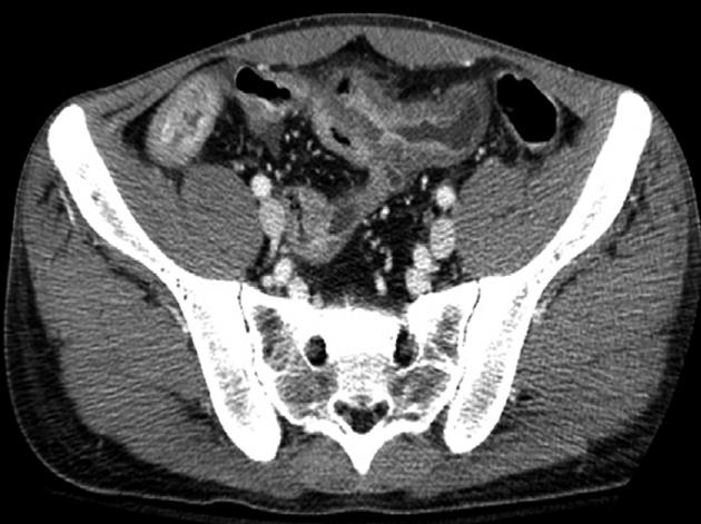
Figure 2 Computed tomography of the abdomen showing evidence of prominent wall thickening with perienteric infiltration in the mid-ileum.
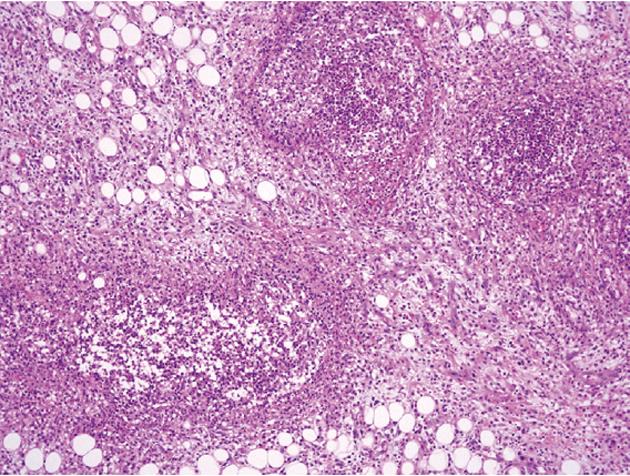
Figure 3 Photography of the pathology, showing the accumulation of many neutrophils in the intravascular lumen (hematoxylin and eosion stain, × 100).
- Citation: Lim JW, Kim KJ, Ye BD, Byeon JS, Myung SJ, Yang SK, Kim JH. Enterovenous fistulization: A rare complication of Crohn’s disease. World J Gastroenterol 2011; 17(47): 5227-5230
- URL: https://www.wjgnet.com/1007-9327/full/v17/i47/5227.htm
- DOI: https://dx.doi.org/10.3748/wjg.v17.i47.5227


