Copyright
©2011 Baishideng Publishing Group Co.
World J Gastroenterol. Dec 7, 2011; 17(45): 5014-5020
Published online Dec 7, 2011. doi: 10.3748/wjg.v17.i45.5014
Published online Dec 7, 2011. doi: 10.3748/wjg.v17.i45.5014
Figure 1 Procedures of experimental study of destruction to porcine spleen in vivo by microwave ablation.
A: The appearance of the spleen before ligation of splenic vein; B: Splenomegalia was seen 2 to 4 h after ligation of the splenic vein; C: The ablation size was measured after microwave ablation.
Figure 2 The ablation lesion size was correlated with the ablation time and power.
A: r = 0.97542, x = 5.5 ± 3.028, y = 23.4 ± 9.023, P < 0.0001; B: r = 0.98258, x = 65.0 ± 24.495, y = 18.47 ± 8.47, P < 0.0001.
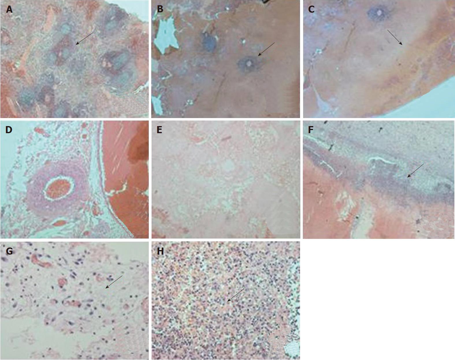
Figure 3 The pathological changes after microwave ablation.
A: Normal spleen tissue (the arrow indicates the splenic corpuscle), hemotoxylin and eosin (HE) × 20; B-D: The pathological changes immediately after microwave ablation (MWA); B: There was some remaining spleen tissue around arterioles in the areas of coagulation necrosis (↑), HE × 20; C: The ablation borderline was not very clear (↑), HE × 20; D: There were no obvious signs of hydropsia or inflammatory reaction, HE × 40; E-H: Pathological changes 1 wk after MWA; E: The coagulation necrosis was well-distributed and complete, HE × 20; F: The ablation borderline was clearer 1 wk after ablation (↑), HE × 20; G: There were some signs of hydropsia and inflammation (↑), HE × 40; H: Many nuclear fragmentations could be seen clearly (↑), HE × 40.
-
Citation: Gao F, Gu YK, Shen JX, Li CL, Jiang XY, Huang JH. Experimental study of destruction to porcine spleen
in vivo by microwave ablation. World J Gastroenterol 2011; 17(45): 5014-5020 - URL: https://www.wjgnet.com/1007-9327/full/v17/i45/5014.htm
- DOI: https://dx.doi.org/10.3748/wjg.v17.i45.5014


