Copyright
©2011 Baishideng Publishing Group Co.
World J Gastroenterol. Nov 28, 2011; 17(44): 4899-4904
Published online Nov 28, 2011. doi: 10.3748/wjg.v17.i44.4899
Published online Nov 28, 2011. doi: 10.3748/wjg.v17.i44.4899
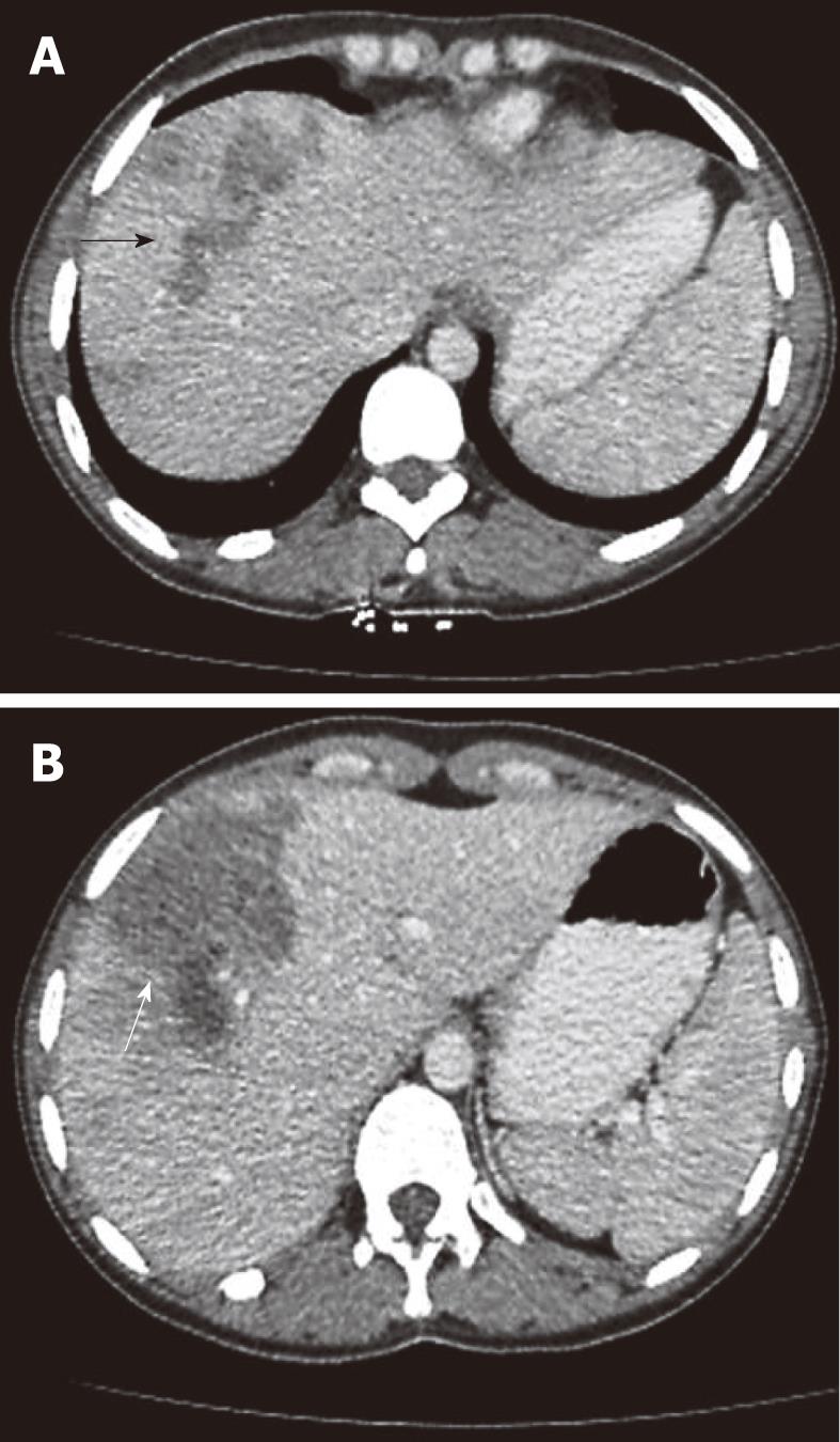
Figure 1 A 30-year-old female patient presented with right upper abdominal pain lasting 8 wk.
A: Tubular branching lesions in the right lobe (arrow); B: Abdominal computerized tomographic examination showed a sub-capsular low density area surrounded by a rim of parenchyma (arrow).
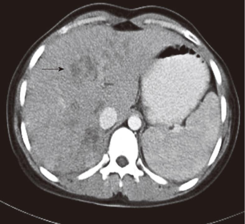
Figure 2 A 19-year-old female patient presented with right upper abdominal pain and fever lasting 3 wk.
Abdominal computerized tomographic examination showed enlargement of the liver and extensive micro-abscesses (arrows).
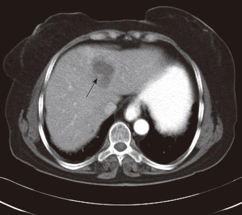
Figure 3 A 70-year-old female patient presented with right upper abdominal pain lasting 16 wk.
Abdominal computerized tomographic examination showed low density masses with hazy margins located to medial segment of the left lobe (arrow).
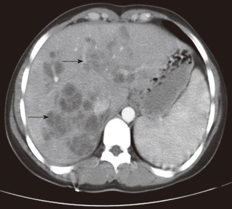
Figure 4 In the patient whose pre-treatment computerized tomographic image is shown in Figure 1, abdominal computerized tomographic examination showed residual lesions (arrow) and minimally enlarged spleen 6 mo after treatment with triclabendazole.
Figure 5 Live and mobil Fasciola hepatica removed from the choledochus by balloon catheter during endoscopic retrograde cholangiopancreatography.
A: A 35-year-old female patient presented with acute pancreatitis associated with elevated liver enzymes. Endoscopic retrograde cholangiopancreatography image showed a radiolucent, roughly crescent-shaped shadow in the common bile duct (arrows); B: A 49-year-old female patient presented with acute cholangitis associated with eosinophilia. Arrow: Head of Fasciola hepatica; Arrowhead: Body of Fasciola hepatica.
- Citation: Kaya M, Beştaş R, Çetin S. Clinical presentation and management of Fasciola hepatica infection: Single-center experience. World J Gastroenterol 2011; 17(44): 4899-4904
- URL: https://www.wjgnet.com/1007-9327/full/v17/i44/4899.htm
- DOI: https://dx.doi.org/10.3748/wjg.v17.i44.4899


