Copyright
©2011 Baishideng Publishing Group Co.
World J Gastroenterol. Nov 21, 2011; 17(43): 4825-4830
Published online Nov 21, 2011. doi: 10.3748/wjg.v17.i43.4825
Published online Nov 21, 2011. doi: 10.3748/wjg.v17.i43.4825
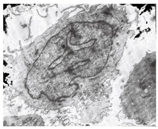
Figure 1 A representative image of monocyte-derive dendritic cells under transmission electron microscope after 7-d induction with recombinant granulocyte-macrophage colony-stimulating factor and interleukin-4 (magnification × 7500).
Dendritic cells were irregular in form, and abundant in extended beard-like prick and mitochondria in cytoplasm.
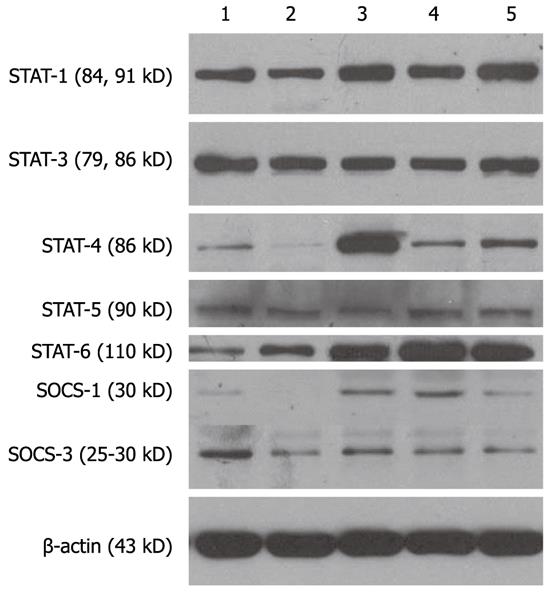
Figure 2 Expression levels of cytoplasmic signal transducers and activators of transcription and suppressors of cell signaling in dendritic cells stimulated with different immunologic adjuvants.
1: Dendritic cells (DCs) from healthy subjects stimulated with synthetic nonmethylated CpG-containing oligodeoxynucleotides (CpG-ODNs); 2-5: DCs from patients with chronic hepatitis B (CHB) stimulated respectively with PBS, CpG-ODNs, TNF-α and CpG-ODNs plus HBsAg. CpG, CpG + HBsAg, TNF-α, and HBsAg are the different adjuvants on DCs. STAT1, 3, 4, 5 and SOCS1, 3 respectively represents the expressing levels of the intracellular signaling molecules including signal transducers and activators of transcription-1, 3, 4, 5 and suppressors of cell signaling-1, 3 in CHB-derived DCs. HBsAg: Hepatitis B surface antigen; TNF: Tumor necrosis factor.
- Citation: Xiang XX, Zhou XQ, Wang JX, Xie Q, Cai X, Yu H, Zhou HJ. Effects of CpG-ODNs on phenotype and function of monocyte-derived dendritic cells in chronic hepatitis B. World J Gastroenterol 2011; 17(43): 4825-4830
- URL: https://www.wjgnet.com/1007-9327/full/v17/i43/4825.htm
- DOI: https://dx.doi.org/10.3748/wjg.v17.i43.4825


