Copyright
©2011 Baishideng Publishing Group Co.
World J Gastroenterol. Nov 14, 2011; 17(42): 4711-4717
Published online Nov 14, 2011. doi: 10.3748/wjg.v17.i42.4711
Published online Nov 14, 2011. doi: 10.3748/wjg.v17.i42.4711
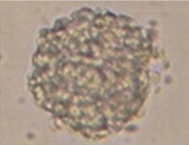
Figure 1 Human colon cancer stem cells grow into clonally derived spheres in serum-free medium.
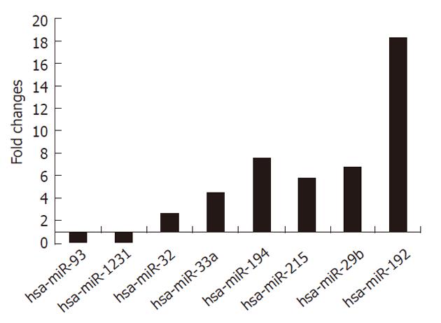
Figure 2 Expression levels of select miRNAs in SW1116csc as measured with quantitative reverse transcription polymerase chain reaction.
Figure 3 Growth curves of SW1116csc, SWcon cells, and SWt cells.
mean ± SD are shown.
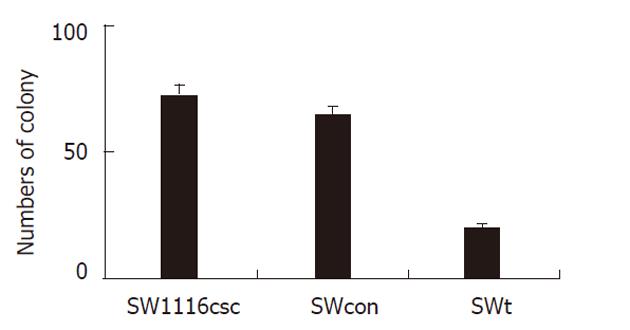
Figure 4 Colony formation after incubation of 100 separate cells for 14 d.
mean ± SD are shown.
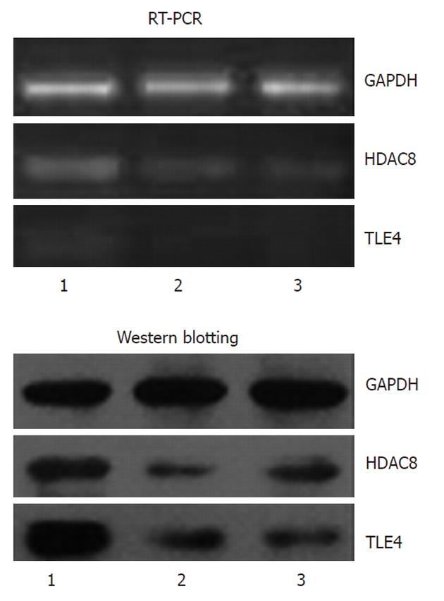
Figure 5 Expression of HDAC8 and TLE4 mRNA and protein in SW1116csc, SW1116 cells and SWt cells.
1: SW1116csc; 2: SW1116 cells; 3: SWt cells. Glyceraldehyde-3-phosphate dehydrogenase was evaluated as an internal control. RT-PCR: Reverse transcription polymerase chain reaction; GAPDH: Glyceraldehyde-3-phosphate dehydrogenase.
- Citation: Yu XF, Zou J, Bao ZJ, Dong J. miR-93 suppresses proliferation and colony formation of human colon cancer stem cells. World J Gastroenterol 2011; 17(42): 4711-4717
- URL: https://www.wjgnet.com/1007-9327/full/v17/i42/4711.htm
- DOI: https://dx.doi.org/10.3748/wjg.v17.i42.4711


