Copyright
©2011 Baishideng Publishing Group Co.
World J Gastroenterol. Nov 14, 2011; 17(42): 4704-4710
Published online Nov 14, 2011. doi: 10.3748/wjg.v17.i42.4704
Published online Nov 14, 2011. doi: 10.3748/wjg.v17.i42.4704
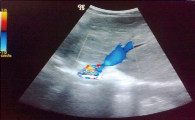
Figure 1 Representative color Doppler ultrasonograph showing a dilated congested left hepatic vein with significant stenosis at the junction thereof with the inferior vena cava.
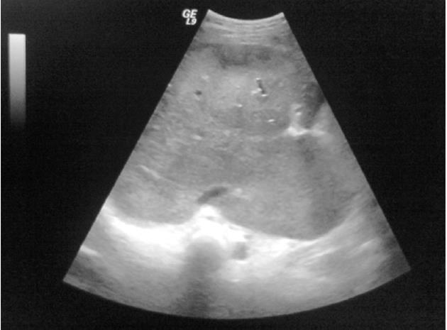
Figure 2 Representative B-mode sonograph showing occlusion of all hepatic veins, a slit-like inferior vena cava and a markedly enlarged caudate lobe.
- Citation: Sakr M, Barakat E, Abdelhakam S, Dabbous H, Yousuf S, Shaker M, Eldorry A. Epidemiological aspects of Budd-Chiari in Egyptian patients: A single-center study. World J Gastroenterol 2011; 17(42): 4704-4710
- URL: https://www.wjgnet.com/1007-9327/full/v17/i42/4704.htm
- DOI: https://dx.doi.org/10.3748/wjg.v17.i42.4704


