Copyright
©2011 Baishideng Publishing Group Co.
World J Gastroenterol. Nov 7, 2011; 17(41): 4619-4624
Published online Nov 7, 2011. doi: 10.3748/wjg.v17.i41.4619
Published online Nov 7, 2011. doi: 10.3748/wjg.v17.i41.4619
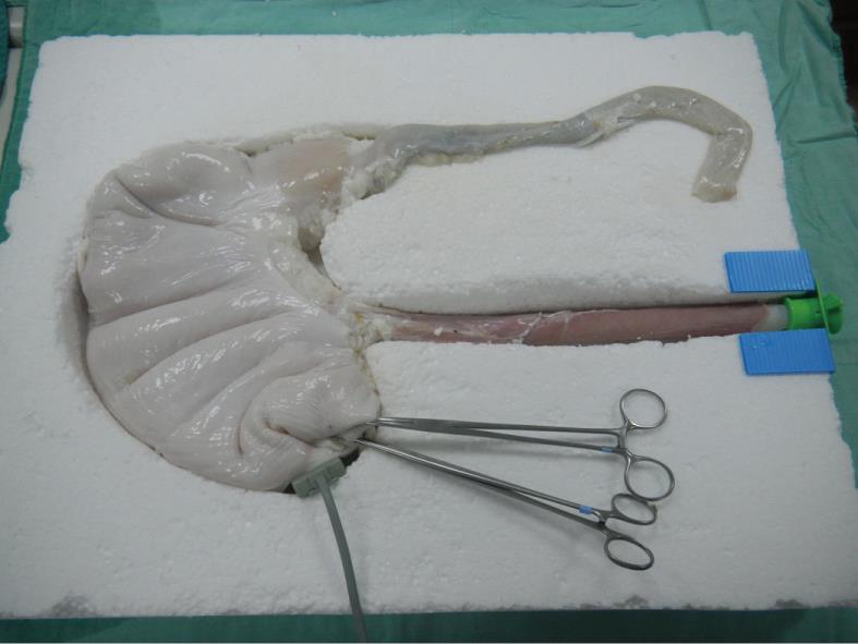
Figure 1 Ex vivo porcine organ package including esophagus, stomach and duodenum in a handmade simulator shell.
Figure 2 Use of the simulator for evaluation.
A: One approximate 8-mm gastric polyp artificially created with a rubber band ligation device in the porcine stomach cardia; B: Snare being advanced to encircle the polyp; C: Successful removal of the entire lesion; D: Application of a hemoclip.
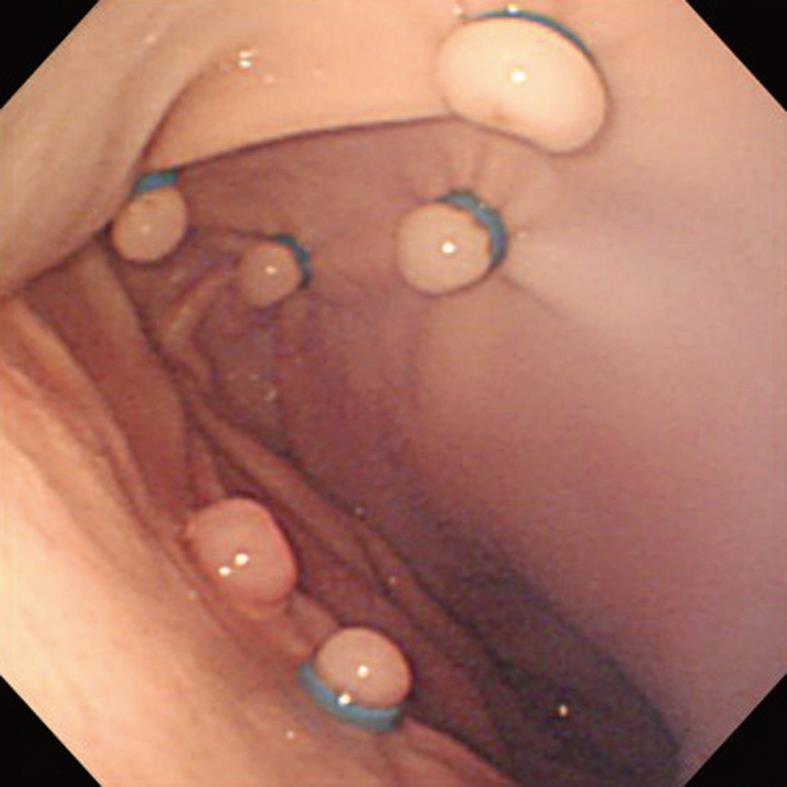
Figure 3 Use of the simulator for training: several artificial polyps placed in different position in the porcine stomach.
Use of a multiband ligation device reduced the time required to load rubber bands, allowing efficient creation of multiple artificial polyps.
-
Citation: Chen MJ, Lin CC, Liu CY, Chen CJ, Chang CW, Chang CW, Lee CW, Shih SC, Wang HY. Training gastroenterology fellows to perform gastric polypectomy using a novel
ex vivo model. World J Gastroenterol 2011; 17(41): 4619-4624 - URL: https://www.wjgnet.com/1007-9327/full/v17/i41/4619.htm
- DOI: https://dx.doi.org/10.3748/wjg.v17.i41.4619


