Copyright
©2011 Baishideng Publishing Group Co.
World J Gastroenterol. Oct 21, 2011; 17(39): 4440-4444
Published online Oct 21, 2011. doi: 10.3748/wjg.v17.i39.4440
Published online Oct 21, 2011. doi: 10.3748/wjg.v17.i39.4440
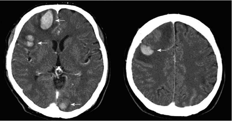
Figure 1 Contrast-enhanced computed tomography scan shows multifocal hemorrhage in the right frontal lobe, right temporal lobe, left occipital lobe, and right parietal lobe (white arrows).
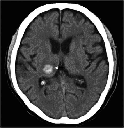
Figure 2 Non-contrast computed tomography scan shows a single right thalamic hemorrhage (white arrow).
- Citation: Nishimura T, Furihata M, Kubo H, Tani M, Agawa S, Setoyama R, Toyoda T. Intracranial hemorrhage in patients treated with bevacizumab: Report of two cases. World J Gastroenterol 2011; 17(39): 4440-4444
- URL: https://www.wjgnet.com/1007-9327/full/v17/i39/4440.htm
- DOI: https://dx.doi.org/10.3748/wjg.v17.i39.4440


