Copyright
©2011 Baishideng Publishing Group Co.
World J Gastroenterol. Oct 21, 2011; 17(39): 4389-4395
Published online Oct 21, 2011. doi: 10.3748/wjg.v17.i39.4389
Published online Oct 21, 2011. doi: 10.3748/wjg.v17.i39.4389
Figure 1 Paris chinensis dioscin inhibits the viability of SGC-7901 cells.
A: Morphological changes of SGC-7901 cells exposed to Paris chinensis dioscin (PCD) for 24 h imaged under a phase contrast microscope at 40 ×; B: Effect of PCD on SGC-7901 viability. SGC-7901 cells were treated with PCD at the indicated concentrations for 0-72 h. Cell viability was then determined by 3-(4,5-dimethylthiazol-2yl)-2,5-diphenyl tetrazolium bromide assay and expressed as the mean ± SD, n = 3. The optical density value at 570 nm is proportional to the number of cells with PCD.
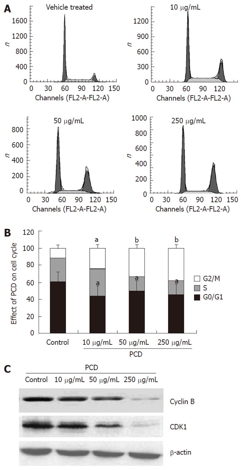
Figure 2 Paris chinensis dioscin induces G2/M cell cycle arrest in SGC-7901 cells.
A: Cell cycle distribution was monitored by flow cytometry using a propidium iodide staining assay; B: Histogram of cell cycle distribution after treatment with Paris chinensis dioscin (PCD) for 24 h. Cell cycle distribution was monitored by flow cytometry using a propidium iodide staining assay. Each histogram represents three parallel experiments, and each bar represents the mean ± SE (One-way ANOVA). aP < 0.05, bP < 0.01 vs vehicle treated (control); C: Western blotting analysis of the expression of cyclin B and CDK1 with or without PCD treatment of SGC-7901 cells.
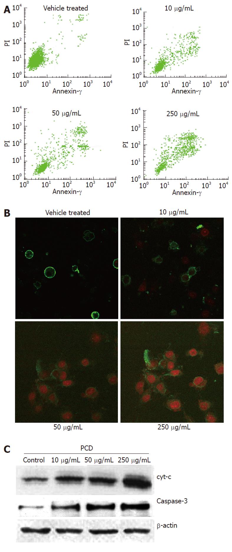
Figure 3 Paris chinensis dioscin induces apoptosis in SGC-7901 cells.
A: Apoptotic cells determined by flow cytometry assay; B: Morphological changes of SGC-7901 cells as determined with a laser scanning confocal microscope at 600 × treated with Paris chinensis dioscin; C: Western blotting analysis of the expressions of caspase-3, cytochrome C and β-actin (internal control) in control and PCD-treated SGC-7901 cells. PCD: Paris chinensis dioscin. PI: Propidium iodide.
Figure 4 Effects of Paris chinensis dioscin on intracellular [Ca2+] expression in human gastric cancer SGC-7901 cells.
A: Fluorescence image of [Ca2+]i under laser scanning confocal microscope at 600 ×; B: Qualitative changes of [Ca2+]i were inferred from the fluorescence intensity after Paris chinensis dioscin (PCD) treatment for 24 h, using SimplePCI imaging systems. Data are presented as mean ± SD (error bar). aP < 0.01 vs control.
-
Citation: Gao LL, Li FR, Jiao P, Yang MF, Zhou XJ, Si YH, Jiang WJ, Zheng TT.
Paris chinensis dioscin induces G2/M cell cycle arrest and apoptosis in human gastric cancer SGC-7901 cells. World J Gastroenterol 2011; 17(39): 4389-4395 - URL: https://www.wjgnet.com/1007-9327/full/v17/i39/4389.htm
- DOI: https://dx.doi.org/10.3748/wjg.v17.i39.4389


