Copyright
©2011 Baishideng Publishing Group Co.
World J Gastroenterol. Oct 14, 2011; 17(38): 4271-4276
Published online Oct 14, 2011. doi: 10.3748/wjg.v17.i38.4271
Published online Oct 14, 2011. doi: 10.3748/wjg.v17.i38.4271
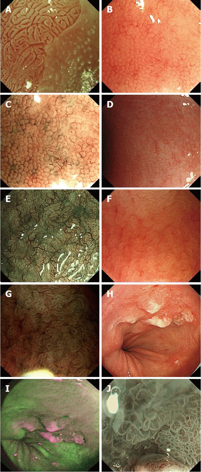
Figure 1 Images of various advanced imaging modalities in Barrett’s oesophagus.
A: Acetic acid used to visualise Barrett’s oesophagus, ridge pattern signifying Intestinal metaplasia; B: High magnification white light endoscopy-round pits in keeping with columnar mucosa without intestinal metaplasia; C: Corresponding area on image B seen with narrow band imaging (NBI) and magnification; D: High magnification white light endoscopy - absent pits in keeping with columnar mucosa with intestinal metaplasia; E: Corresponding area on image D seen with NBI and magnification; F: High magnification white light endoscopy - villous/ridge pits in keeping with columnar mucosa with intestinal metaplasia; G: Corresponding area on image F seen with NBI and magnification; H: White light endoscopy of Barrett’s cancer; I: Corresponding area on autoflourescence imaging; J: Abnormal area on NBI with magnification showing total distortion of the pit pattern.
- Citation: Singh R, Mei SCY, Sethi S. Advanced endoscopic imaging in Barrett's oesophagus: A review on current practice. World J Gastroenterol 2011; 17(38): 4271-4276
- URL: https://www.wjgnet.com/1007-9327/full/v17/i38/4271.htm
- DOI: https://dx.doi.org/10.3748/wjg.v17.i38.4271


