Copyright
©2011 Baishideng Publishing Group Co.
World J Gastroenterol. Sep 21, 2011; 17(35): 4044-4047
Published online Sep 21, 2011. doi: 10.3748/wjg.v17.i35.4044
Published online Sep 21, 2011. doi: 10.3748/wjg.v17.i35.4044
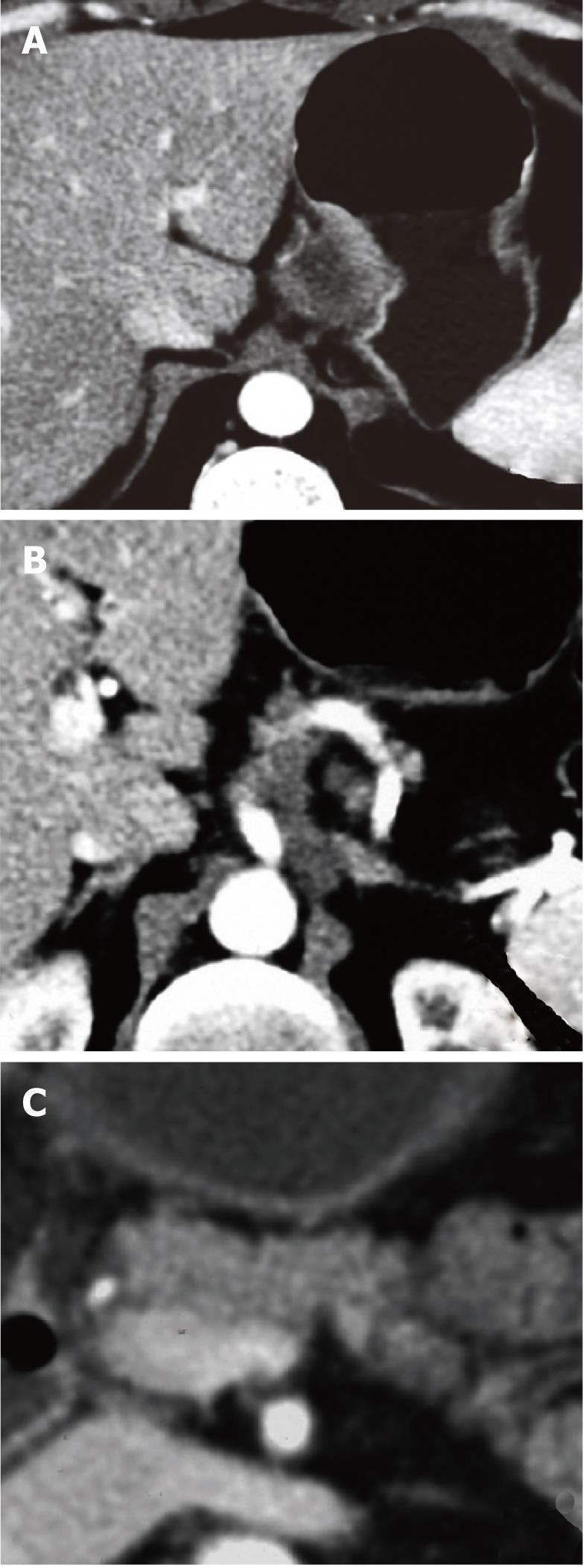
Figure 1 Contrast-enhanced computed tomography scan.
A cardia tumor is evident (A), associated with multiple enlarged nodes infiltrating the celiac trunk origin (B) and the pancreatic body (C).
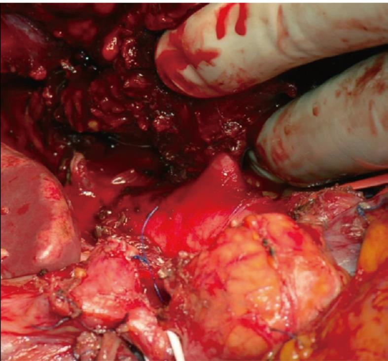
Figure 2 Intraoperative findings.
Celiac trunk origin from the aorta is free from tumor infiltration.
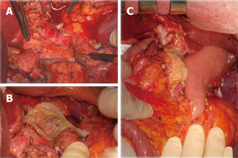
Figure 3 Intraoperative findings after the demolitive phase.
A: From the left, the distal hepatic artery stump, the celiac stump and the superior mesenteric artery; B, C: Tachosil® is then applied over the aorta (B) and over the pancreas stump (C).
- Citation: Baiocchi G, Portolani N, Gheza F, Giulini SM. Collagen-based biological glue after Appleby operation for advanced gastric cancer. World J Gastroenterol 2011; 17(35): 4044-4047
- URL: https://www.wjgnet.com/1007-9327/full/v17/i35/4044.htm
- DOI: https://dx.doi.org/10.3748/wjg.v17.i35.4044


