Copyright
©2011 Baishideng Publishing Group Co.
World J Gastroenterol. Sep 21, 2011; 17(35): 4023-4030
Published online Sep 21, 2011. doi: 10.3748/wjg.v17.i35.4023
Published online Sep 21, 2011. doi: 10.3748/wjg.v17.i35.4023
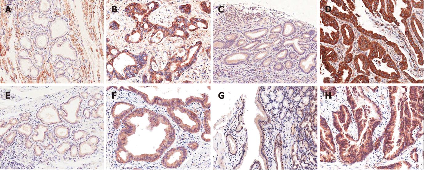
Figure 1 Notch receptor expression in cholangiocarcinomas (Immunohistochemistry).
A, E: Notch receptor 1; B, F: Notch receptor 2; C, G: Notch receptor 3; D, H: Notch receptor 4; A-D: Non-neoplastic tissue (x 40); E-H: Cholangiocarcinomas (CCS) (x 100). Non-neoplastic biliary glandular or surface epithelial cells show weak cytoplasmic staining. High grade expression of Notch receptors is detected in cytoplasm of cancer cells of CCs.
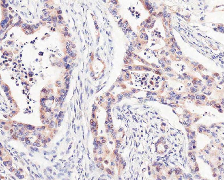
Figure 2 Cytoplasmonuclear localization of Notch receptor 3 in moderately differentiated cholangiocarcinoma (immunohistochemistry, × 200).
Brown-colored positive immunostaining of cholangiocarcinoma cells in both cytoplasm and nuclei is frequently observed.
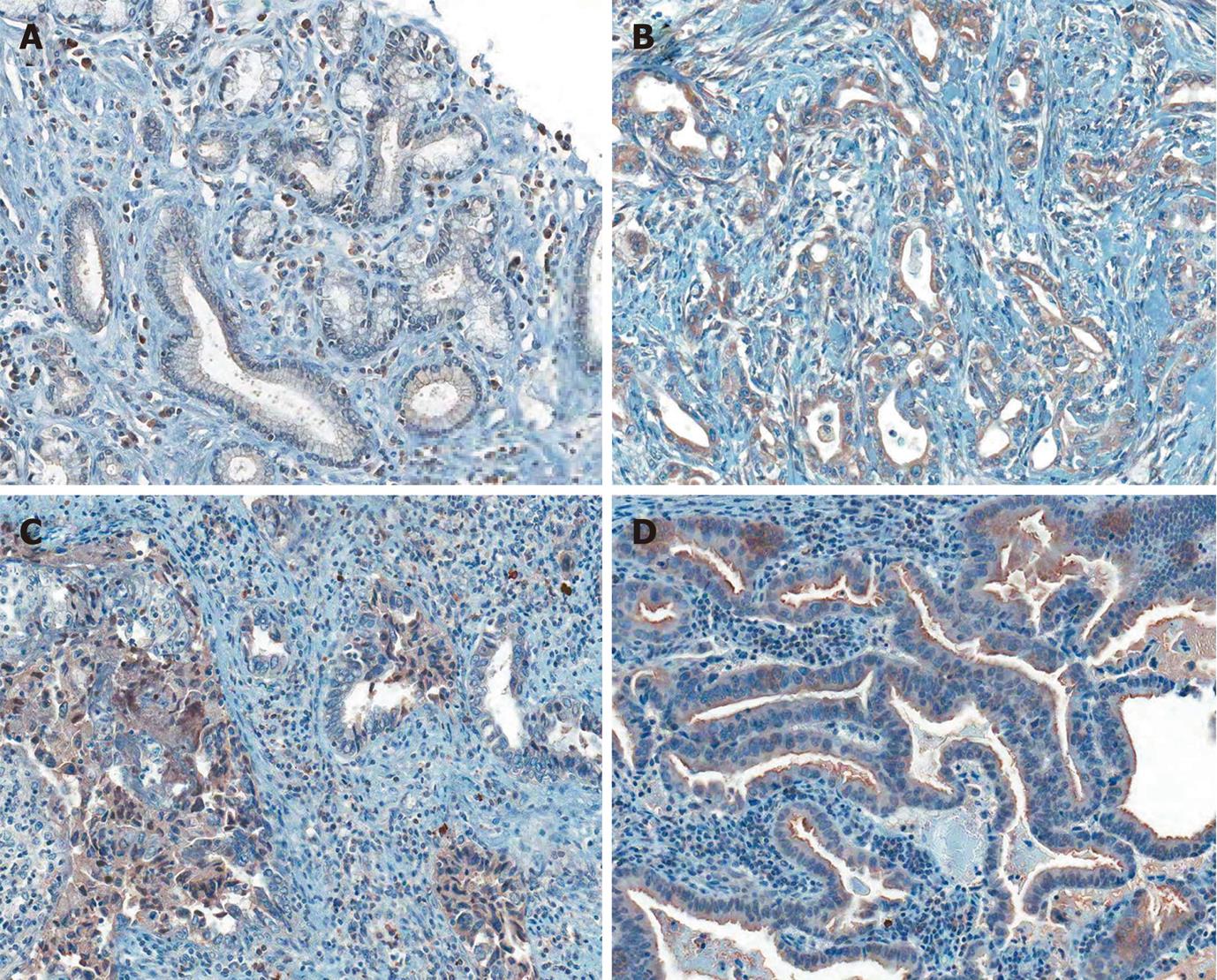
Figure 3 Delta-like ligand-4 expression in cholangiocarcinomas (immunohistochemistry, × 100).
A: Non-neoplastic biliary tissue; B-D: Cholangiocarcinomas. Non-neoplastic biliary epithelial cells (weak) and stromal inflammatory cells (strong) show cytoplasmic staining. Brown-colored expression of delta-like ligand-4 in cholangiocarcinoma cells is located at cytoplasm (B-D), coexisting nuclei (C), and luminal border (D).
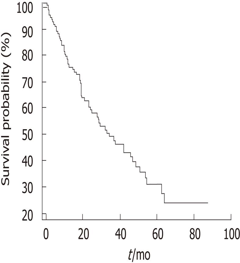
Figure 4 Overall survival curve using the Kaplan-Meier method by log rank test.
Median survival is 34.1 mo.
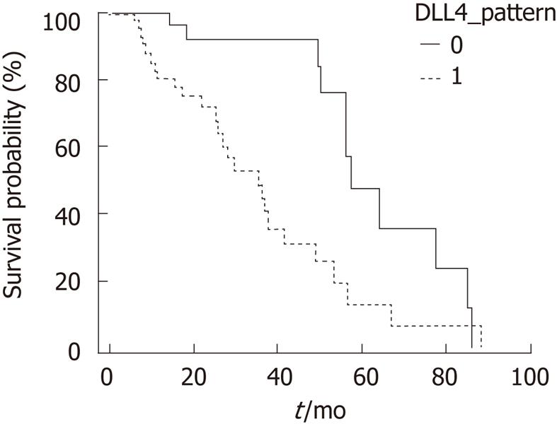
Figure 5 Survival curves of Cholangiocarcinomas with Delta-like ligand-4 expression with or without coexistent cytoplasmonuclear localization using the Kaplan-Meier method by log rank test.
0: Without nuclear localization; 1: With coexistent cytoplasmonuclear localization. Cases of coexistent cytoplasmonuclear localization of Delta-like ligand-4 expression show poor survival (P = 0.002).
- Citation: Yoon HA, Noh MH, Kim BG, Han JS, Jang JS, Choi SR, Jeong JS, Chun JH. Clinicopathological significance of altered Notch signaling in extrahepatic cholangiocarcinoma and gallbladder carcinoma. World J Gastroenterol 2011; 17(35): 4023-4030
- URL: https://www.wjgnet.com/1007-9327/full/v17/i35/4023.htm
- DOI: https://dx.doi.org/10.3748/wjg.v17.i35.4023


