Copyright
©2011 Baishideng Publishing Group Co.
World J Gastroenterol. Apr 21, 2011; 17(15): 2037-2043
Published online Apr 21, 2011. doi: 10.3748/wjg.v17.i15.2037
Published online Apr 21, 2011. doi: 10.3748/wjg.v17.i15.2037
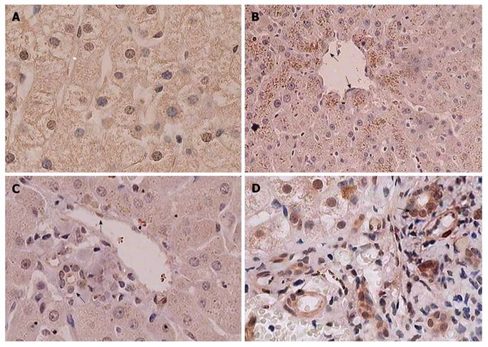
Figure 1 Differentiated embryo chondrocyte 1 expression in normal liver tissue by immunohistochemical staining.
A: Normal liver tissues showing strong nuclear differentiated embryo chondrocyte 1 (DEC1) staining and diffuse cytoplasmic staining in hepatocytes, × 400; B: Strong granular staining of DEC1 in cytoplasm of hepatocytes around the central vein in the intact hepatic lobule, × 200; C: Absent granular staining and diffuse DEC1 staining pattern around portal area, × 400; D: Liver mesenchymal tissues, × 400. Endothelial cells (single arrow) and bile duct epithelial cells (double arrow) showing cytoplasm and/or nuclear DEC1 immunoreactivity.
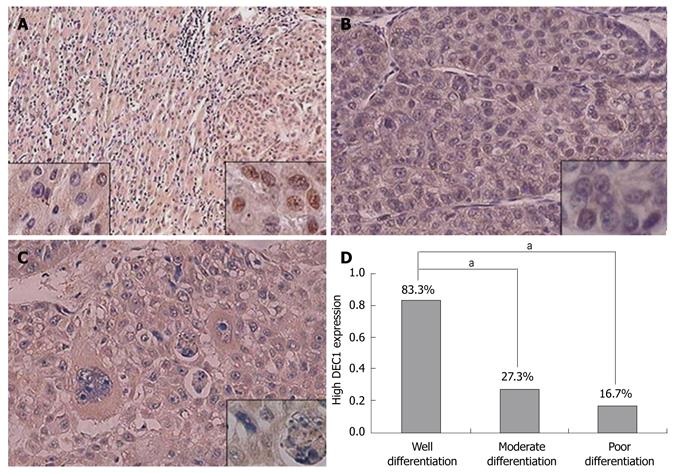
Figure 2 Differentiated embryo chondrocyte 1 expression in hepatocellular carcinoma by immunohistochemical staining.
A: Well-differentiated hepatocellular carcinoma (HCC), × 100. Intense nuclear staining in HCC (right inset), and weak nuclear staining in the corresponding adjacent non-tumor tissues (left inset), with no significant difference in cytoplasmic staining; B: Moderately-differentiated HCC, × 200; C: Poorly-differentiated HCC, × 200. Inserted figure (at high magnification) showing differentiated embryo chondrocyte 1 (DEC1) staining in the cytoplasm and nucleus, × 400; D: Quantitation of DEC1 nuclear expression in HCC by different differentiation status. aP < 0.05.
- Citation: Shi XH, Zheng Y, Sun Q, Cui J, Liu QH, Qü F, Wang YS. DEC1 nuclear expression: A marker of differentiation grade in hepatocellular carcinoma. World J Gastroenterol 2011; 17(15): 2037-2043
- URL: https://www.wjgnet.com/1007-9327/full/v17/i15/2037.htm
- DOI: https://dx.doi.org/10.3748/wjg.v17.i15.2037


