Copyright
©2011 Baishideng Publishing Group Co.
World J Gastroenterol. Apr 21, 2011; 17(15): 1961-1970
Published online Apr 21, 2011. doi: 10.3748/wjg.v17.i15.1961
Published online Apr 21, 2011. doi: 10.3748/wjg.v17.i15.1961
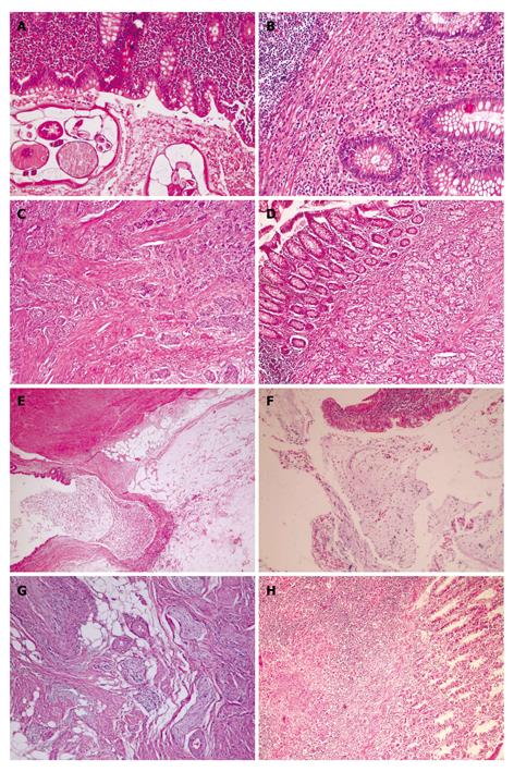
Figure 1 Unusual histopathologic findings.
A: Adult of E.Vermicularis in appendices (HE, × 200); B: Eosinophilic appendicitis: diffuse eosinophilic infiltrate in lamina propria (HE, × 200); C: Carcinoid tumor of classic type is formed by solid nest of small monotonous cells with occasional acinar formation (HE, × 100); D: Microglandular goblet cell carcinoma. Acute appendicitis with a diffusely infiltrating goblet cell neoplasm. tumor cells infiltrated muscularis propria (HE, × 200); E: Mucosel. Dilatation of lumen by mucinous secretion, thin appendiceal wall. Mucin is protruding into surrounding fatty tissue (HE, × 40); F: Mucinous cystadenoma of appendix. Typical epithelium of a cystadenoma with pseudostratified, columnar cells containing elonged, crowded, hyperchromatic nuclei and scattered goblet cells with mucus in cavity (HE, × 100); G: Neurogenous hyperplasia of appendix. The proliferating spindle cells shown in this photography (HE, × 200); H: Tuberculous appendicitis. Granuloma which contain a caseating center surrounded by epithelioid cells, lymphocytes and histiocytes. A giant cell is present in the granuloma (HE, × 20).
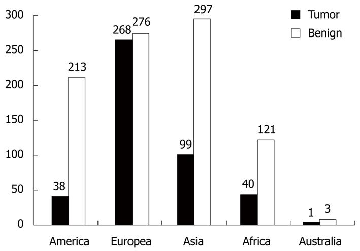
Figure 2 Worldwide distribution of the 1366 cases defined as ''unusual findings''.
Tumor: Carcinoid, goblet cell carcinoid, mucocele, appendix adenocarcinoma, lymphoma, mucinous cystadenoma and adenocarcinoma, polypoid lesions, leukemia, gastrointestinal stromal tumor, dysplastic change; Benign: Non-tumoral causes.
- Citation: Akbulut S, Tas M, Sogutcu N, Arikanoglu Z, Basbug M, Ulku A, Semur H, Yagmur Y. Unusual histopathological findings in appendectomy specimens: A retrospective analysis and literature review. World J Gastroenterol 2011; 17(15): 1961-1970
- URL: https://www.wjgnet.com/1007-9327/full/v17/i15/1961.htm
- DOI: https://dx.doi.org/10.3748/wjg.v17.i15.1961


