Copyright
©2011 Baishideng Publishing Group Co.
World J Gastroenterol. Apr 14, 2011; 17(14): 1923-1926
Published online Apr 14, 2011. doi: 10.3748/wjg.v17.i14.1923
Published online Apr 14, 2011. doi: 10.3748/wjg.v17.i14.1923
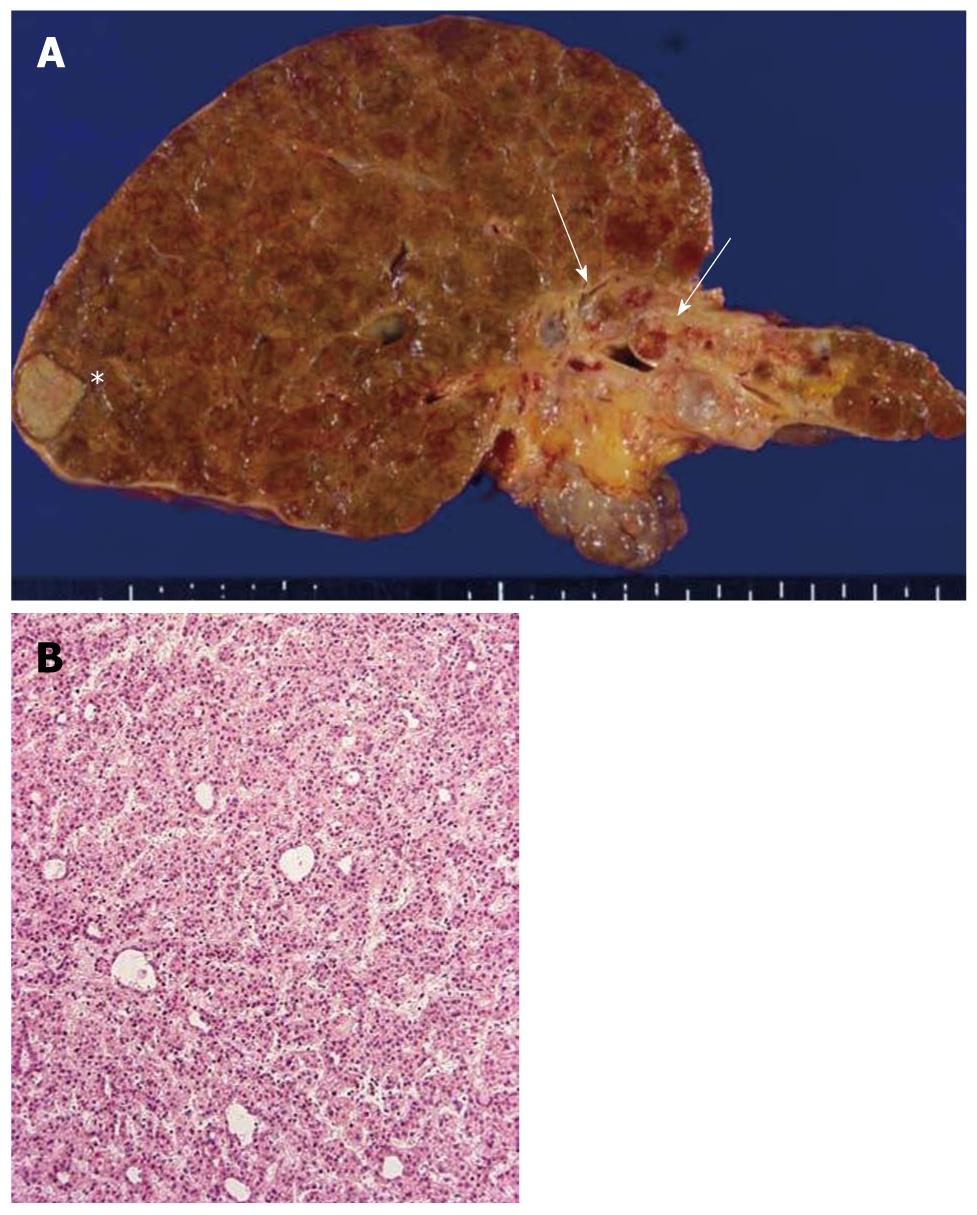
Figure 1 Liver pathology.
A: The autopsied liver tissue shows cirrhosis with a solid whitish lesion (*) in segment 6 of the right lobe (hepatocellular carcinoma after transcatheter arterial chemoembolization) and cystic dilatation in the left intrahepatic bile ducts filling with yellowish-tan papillary masses and mucin production (arrows). The left liver appears atrophied; B: Well-differentiated hepatocellular carcinoma. Hematoxylin and eosin staining (H and E).
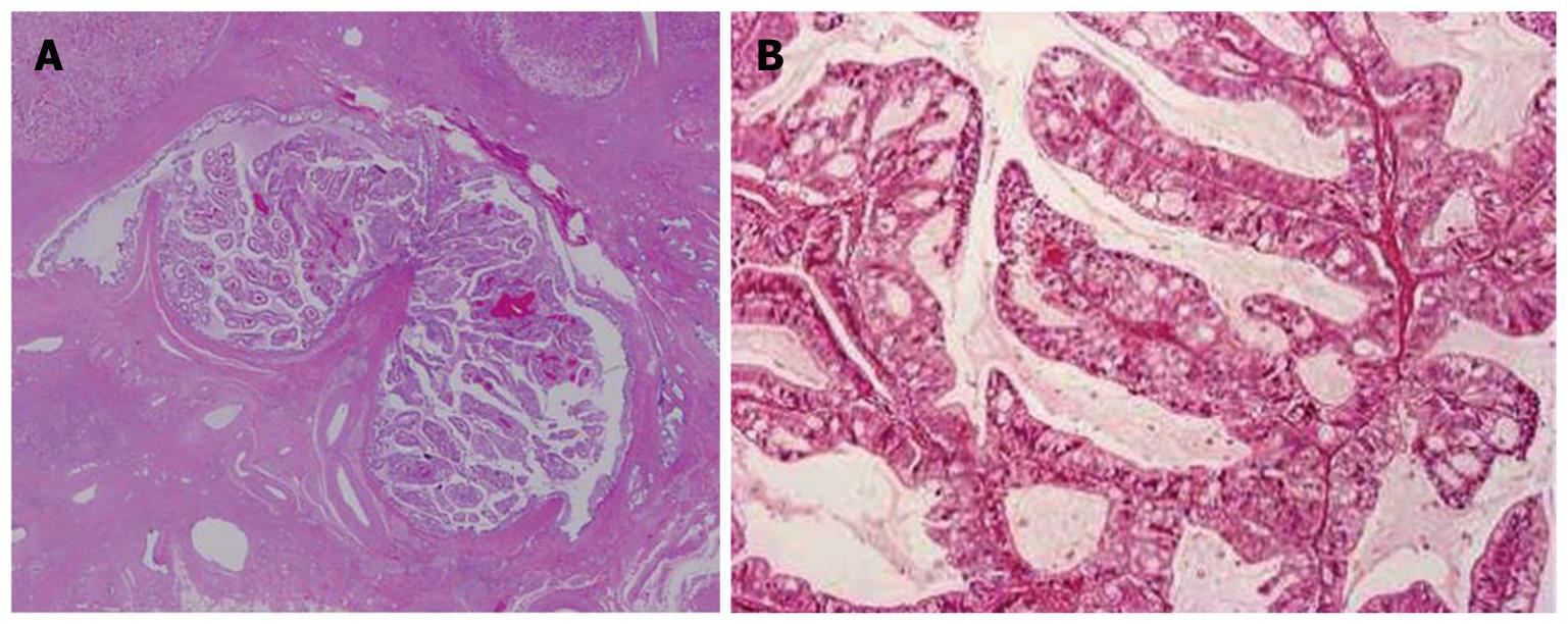
Figure 2 Histological features of the papillary tumor of the left lobe.
A: Neoplastic biliary epithelia show intraductal papillary growth in the dilated bile duct lumen. Hematoxylin and eosin staining (H and E); B: The atypical biliary epithelium with a fine fibrovascular core is spreading. H and E.
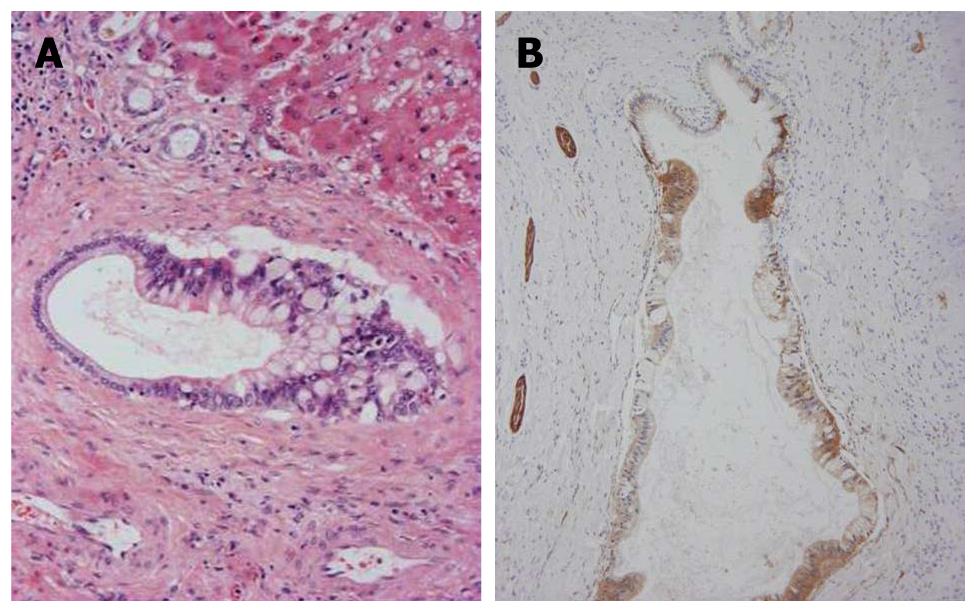
Figure 3 Carcinomatouscholangiocytes.
A: Atypical biliary epithelial cells (cholangiocarcinoma) partially replace the epithelia of septal bile ducts. Hematoxylin and eosin staining (H and E); B: Neoplastic biliary epithelial cells spreading on the luminal surface of the septal bile ducts are positive for neural cell adhesion molecule (NCAM). Nerve fibers around the bile ducts are also positive. Immunostaining for NCAM and hematoxylin.
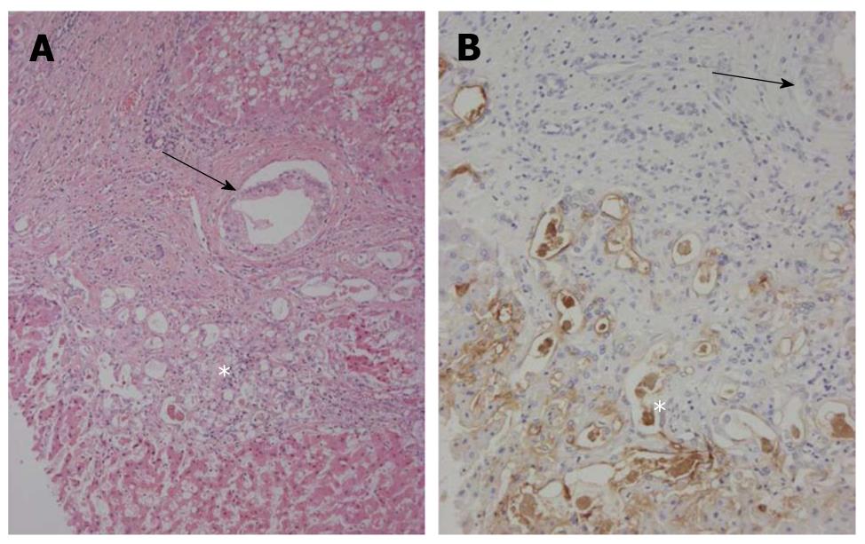
Figure 4 Bileductular cell proliferation.
A: Clusters of atypical bile ductular cells (*) are seen, and are regarded as malignant cells. An arrow shows the involvement of carcinoma cells of interlobular bile ducts. Hematoxylin and eosin staining (H and E); B: Atypical bile ductular cells (*) are positive for mucin (MUC)1. Neoplastic epithelial cells replacing interlobular bile ducts (arrow) are also focally positive. Immunostaining for MUC1 and hematoxylin.
- Citation: Xu J, Sato Y, Harada K, NorihideYoneda, Ueda T, Kawashima A, AkishiOoi, YasuniNakanuma. Intraductal papillary neoplasm of the bile duct in liver cirrhosis with hepatocellular carcinoma. World J Gastroenterol 2011; 17(14): 1923-1926
- URL: https://www.wjgnet.com/1007-9327/full/v17/i14/1923.htm
- DOI: https://dx.doi.org/10.3748/wjg.v17.i14.1923


