Copyright
©2011 Baishideng Publishing Group Co.
World J Gastroenterol. Apr 14, 2011; 17(14): 1915-1922
Published online Apr 14, 2011. doi: 10.3748/wjg.v17.i14.1915
Published online Apr 14, 2011. doi: 10.3748/wjg.v17.i14.1915
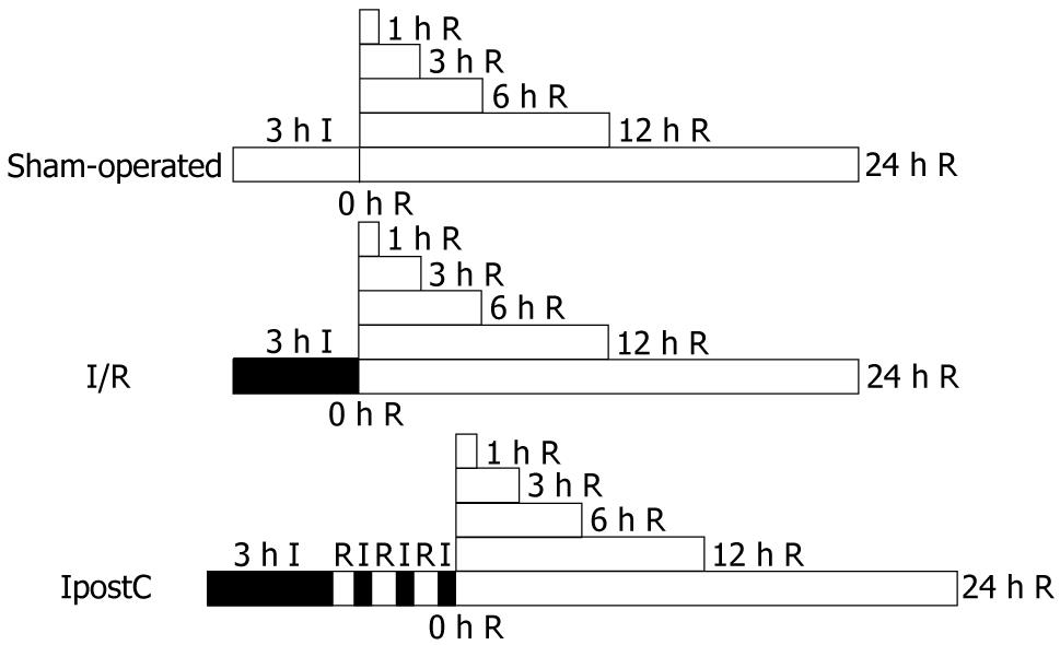
Figure 1 Experimental protocol.
In the sham-operated group (n = 36) there was no intervention; ischemic/reperfusion (I/R, n = 36) was elicited by 3 h I followed by 0, 1, 3, 6, 12 or 24 h R; ischemic post-conditioning (IpostC) (n = 36) was performed by 3 circles of 30 s of R followed by 30 s of I before 0, 1, 3, 6, 12 or 24 h of R, respectively. R: reperfusion; I: ischemia.
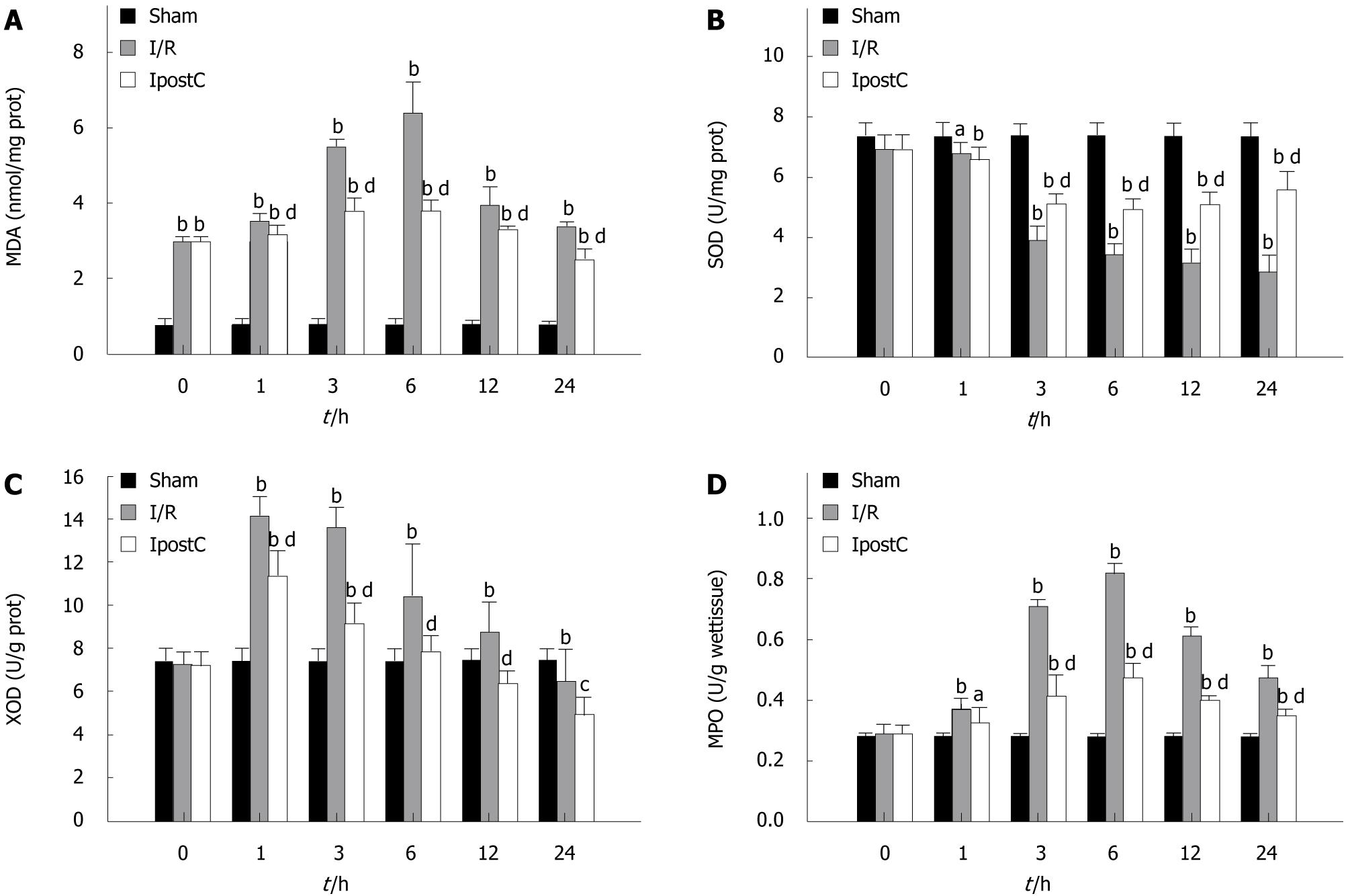
Figure 2 Gastric oxidative stress and lipid peroxidation after gastric ischemia/reperfusion injury of 3 h of ischemia and 24 h of reperfusion (mean ± SE, n = 36).
A: The level of malondialdehyde (MDA); B: The activity of superoxide dismutase (SOD); C: The activity of xanthine oxidase (XOD); D: The activity of myeloperoxidase (MPO). The activity of SOD decreased with the activities of XOD, MPO and the level of MDA increased in the ischemia/reperfusion (I/R) group compared with those of the sham-operated group at 1, 3, 6, 12, and 24 h. However, ischemic post-conditioning (IpostC) treatment significantly increased the activity of SOD, and decreased the activities of XOD, MPO and the level of MDA at each time. aP< 0.05, bP< 0.01 vs sham group; cP< 0.05, dP< 0.01 vs I/R group.
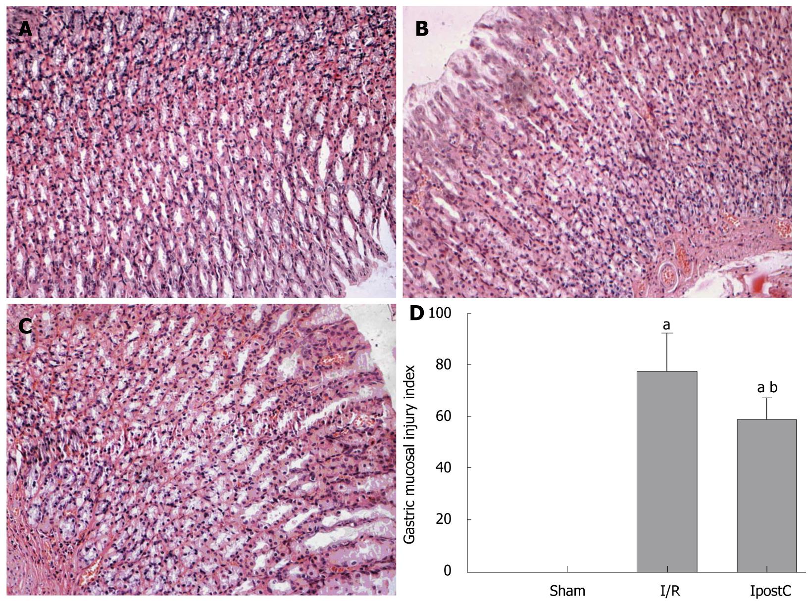
Figure 3 Histological evaluations of gastric tissue.
Representative gastric sections were obtained 6 h after sham-operated surgery or ischemia/reperfusion (I/R). A: Section from sham-operated group; B: Section from I/R group; C: Section from IpostC group. All of the sections stained with hema-toxylin and eosin × 200; D: Yong-Mei Zhang scores for acute gastric lesions from sham, I/R and ischemic post-conditioning (IpostC) groups (mean ± SD; n = 6); aP< 0.01 vs sham-operated group; bP< 0.01 vs I/R group.
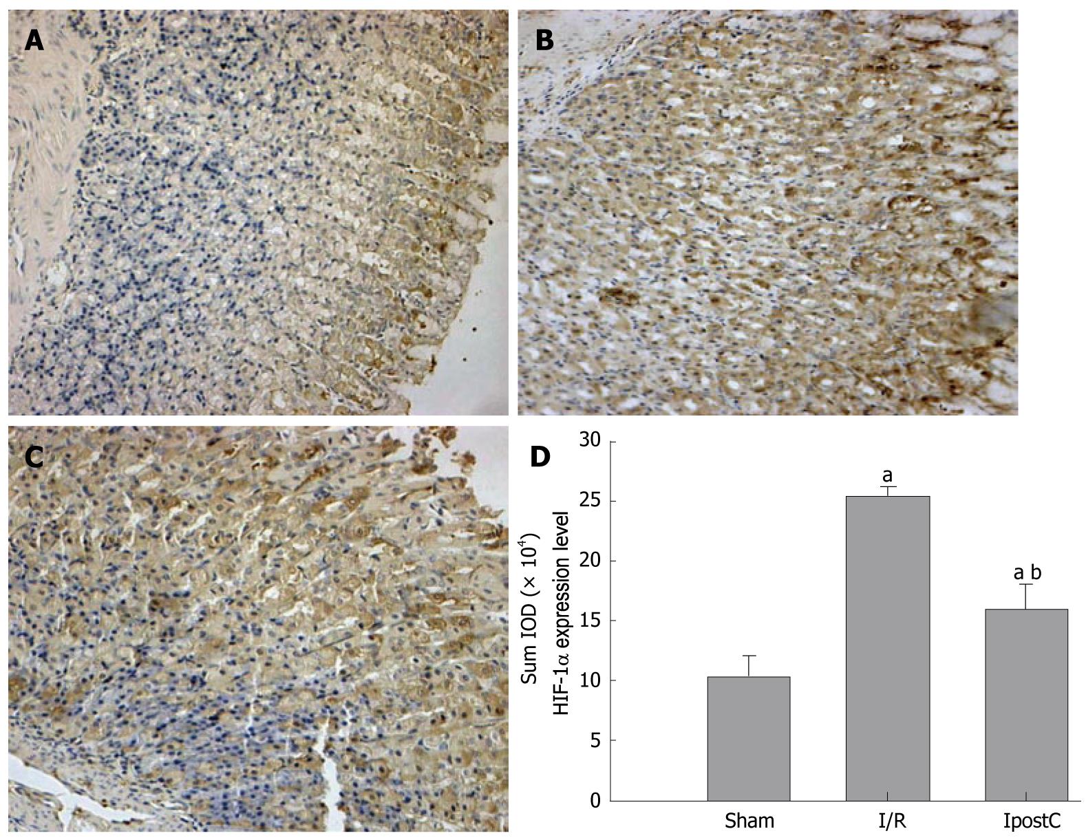
Figure 4 Photomicrographs of hypoxia-induced factor 1α (HIF-1α) immunohistochemistry in the gastric tissue (magnification 20 ×).
The thickness of gastric containing HIF-1α-positive gastric cells was determined at a 6-h time point. A: Weak the glandular epithelial cytoplasm and the vascular endothelial cytoplasm staining in Sham-operated group gastric; B: Strong the glandular epithelial cytoplasm, and the vascular endothelial cytoplasm and nuclear staining in ischemia/reperfusion (I/R) group gastric; C: Moderate the glandular epithelial cytoplasm, and the vascular endothelial cytoplasm and nuclear staining in ischemic post-conditioning (IpostC) group gastric; D: SUM IOD of HIF-1α expression level in sham-operated, I/R and IpostC groups of gastric tissue (mean ± SD; n < 8); aP< 0.01 vs sham-operated group; bP< 0.01 vs I/R group.
- Citation: Wang T, Leng YF, Zhang Y, Xue X, Kang YQ, Zhang Y. Oxidative stress and hypoxia-induced factor 1α expression in gastric ischemia. World J Gastroenterol 2011; 17(14): 1915-1922
- URL: https://www.wjgnet.com/1007-9327/full/v17/i14/1915.htm
- DOI: https://dx.doi.org/10.3748/wjg.v17.i14.1915


