Copyright
©2011 Baishideng Publishing Group Co.
World J Gastroenterol. Mar 21, 2011; 17(11): 1501-1506
Published online Mar 21, 2011. doi: 10.3748/wjg.v17.i11.1501
Published online Mar 21, 2011. doi: 10.3748/wjg.v17.i11.1501
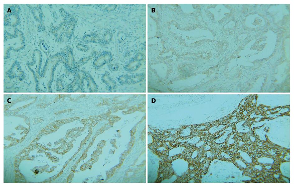
Figure 1 Immunohistochemistry staining of gastric carcinoma tissue samples showing negative HER-2 protein expression (A, B), positive HER-2 protein expression (C, D) (original magnification × 200).
Figure 2 Fluorescence in situ hybridization targeting HER-2 in gastric carcinoma specimens showing intestinal type of carcinoma without HER-2 gene amplification (A) and intestinal type of carcinoma with HER-2 gene amplification (B) (original magnification × 1000).
- Citation: Yan SY, Hu Y, Fan JG, Tao GQ, Lu YM, Cai X, Yu BH, Du YQ. Clinicopathologic significance of HER-2/neu protein expression and gene amplification in gastric carcinoma. World J Gastroenterol 2011; 17(11): 1501-1506
- URL: https://www.wjgnet.com/1007-9327/full/v17/i11/1501.htm
- DOI: https://dx.doi.org/10.3748/wjg.v17.i11.1501


