Copyright
©2011 Baishideng Publishing Group Co.
World J Gastroenterol. Jan 7, 2011; 17(1): 63-68
Published online Jan 7, 2011. doi: 10.3748/wjg.v17.i1.63
Published online Jan 7, 2011. doi: 10.3748/wjg.v17.i1.63
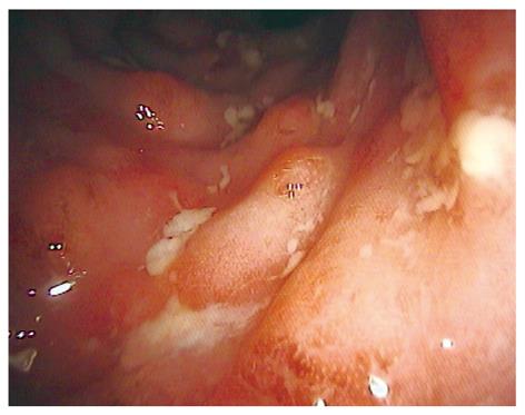
Figure 1 High-resolution standard white light endoscopic image of active Crohn’s disease.
Endoscopy shows ulcerations, mucosal edema and erythema.
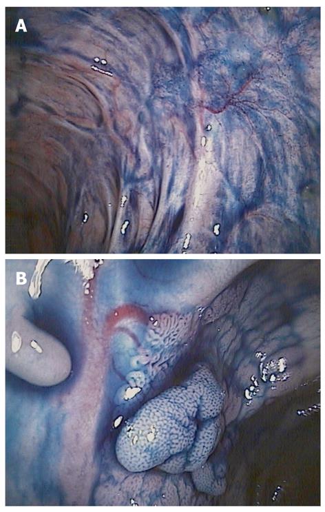
Figure 2 Chromoendoscopy with indigo carmine.
A better distinction of mucosal changes in long standing ulcerative colitis (A) and pit pattern analysis of suspicious lesions (B).
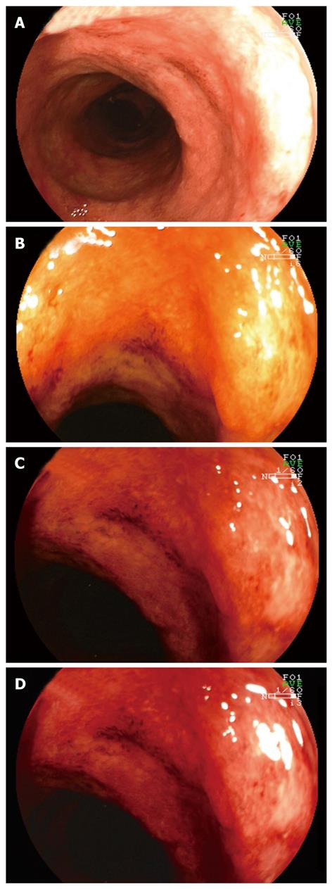
Figure 3 Virtual chromoendoscopy using the fujinon intelligent color enhancement-system.
A: Shows standard white light endoscopic image; B-D: Illustrate different fujinon intelligent color enhancement settings to improve mucosal detail.
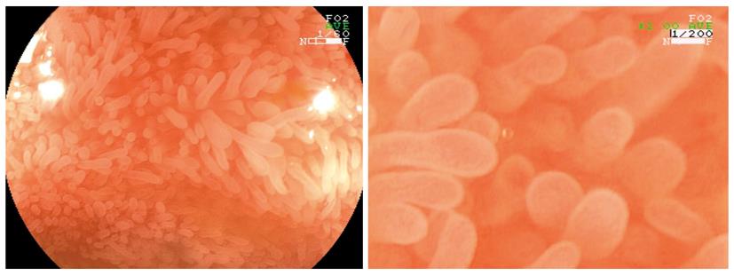
Figure 4 High-magnification endoscopy of ileum mucosa in patient with Crohn’s disease without activity.
Villi are clearly visualized.
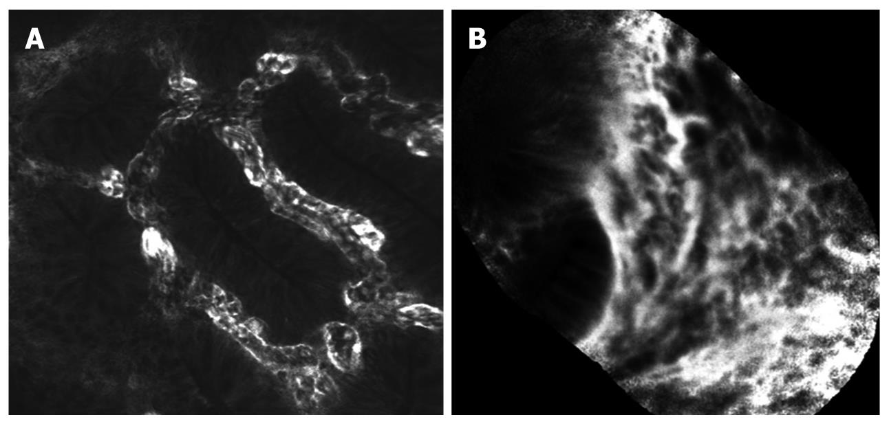
Figure 5 Confocal laser endomicroscopy using either the integrated system (iCLE, A) or the probe-based system (pCLE, B) visualizes dilated microvessels, leakage and disturbed crypt architecture in active ulcerative colitis.
Figure 6 Endocytoscopy enables visualization of different cytological and architectural features, including size, arrangement, and density of cells.
- Citation: Neumann H, Neurath MF, Mudter J. New endoscopic approaches in IBD. World J Gastroenterol 2011; 17(1): 63-68
- URL: https://www.wjgnet.com/1007-9327/full/v17/i1/63.htm
- DOI: https://dx.doi.org/10.3748/wjg.v17.i1.63


