Copyright
©2011 Baishideng Publishing Group Co.
World J Gastroenterol. Jan 7, 2011; 17(1): 42-48
Published online Jan 7, 2011. doi: 10.3748/wjg.v17.i1.42
Published online Jan 7, 2011. doi: 10.3748/wjg.v17.i1.42
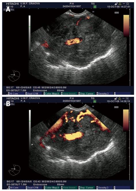
Figure 1 Contrast enhanced endoscopic ultrasonography exam of lung adenocarcinoma.
A: Non-enhanced power Doppler image of a lung adenocarcinoma visualized in the aorto-pulmonary window from the mid-esophagus, with discrete Doppler signals in the periphery of the mass and embedding of a large branch of the left pulmonary artery; B: Same tumor visualized after contrast-enhancement with SonoVue, with a better depiction of the vascular peripheral signals and the possibility of quantification of the vascular index. The relationship to the aorta and pulmonary artery is clearly depicted.
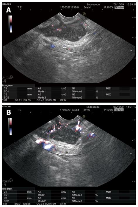
Figure 2 Contrast enhanced endoscopic ultrasonography of pancreatic cancer.
A: Pancreatic head adenocarcinoma visualized in bidirectional non-enhanced power Doppler mode; B: Contrast-enhancement with SonoVue indicates a hypovascular mass with increased collateral circulation.
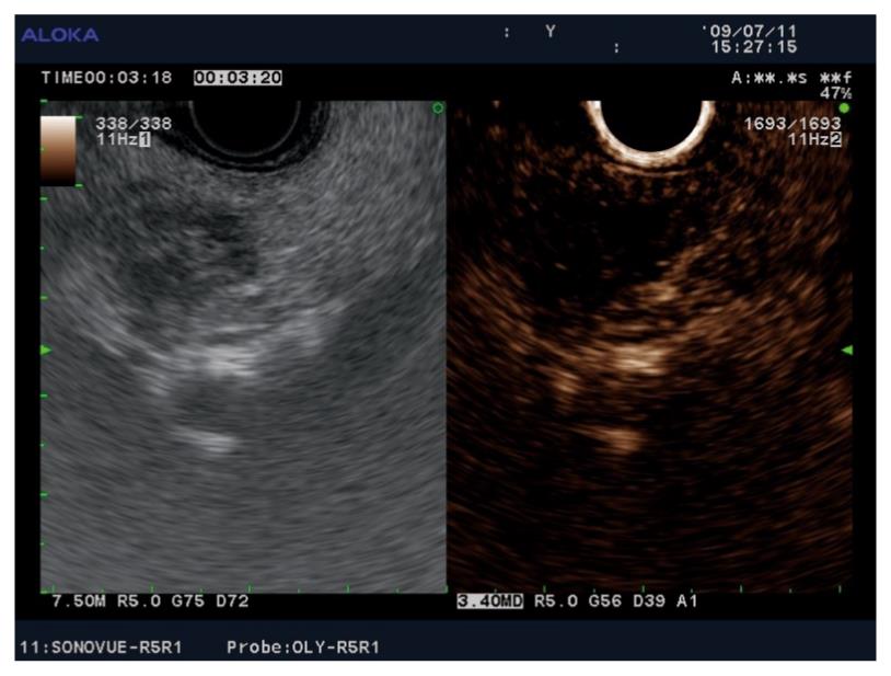
Figure 3 Contrast-enhanced (SonoVue) harmonic endoscopic ultrasound imaging showing a small (12 mm) hypovascular adenocarcinoma in the head of the pancreas.
The tumor tissue did not enhance in the early arterial phase, nor in the late venous phase, as compared to the surrounding pancreatic parenchyma.
- Citation: Reddy NK, Ioncică AM, Săftoiu A, Vilmann P, Bhutani MS. Contrast-enhanced endoscopic ultrasonography. World J Gastroenterol 2011; 17(1): 42-48
- URL: https://www.wjgnet.com/1007-9327/full/v17/i1/42.htm
- DOI: https://dx.doi.org/10.3748/wjg.v17.i1.42


