
















©2011 Baishideng Publishing Group Co.
World J Gastroenterol. Jan 7, 2011; 17(1): 42-48
Published online Jan 7, 2011. doi: 10.3748/wjg.v17.i1.42
Published online Jan 7, 2011. doi: 10.3748/wjg.v17.i1.42
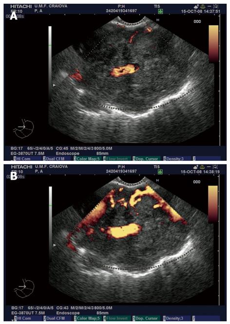
Figure 1 Contrast enhanced endoscopic ultrasonography exam of lung adenocarcinoma.
A: Non-enhanced power Doppler image of a lung adenocarcinoma visualized in the aorto-pulmonary window from the mid-esophagus, with discrete Doppler signals in the periphery of the mass and embedding of a large branch of the left pulmonary artery; B: Same tumor visualized after contrast-enhancement with SonoVue, with a better depiction of the vascular peripheral signals and the possibility of quantification of the vascular index. The relationship to the aorta and pulmonary artery is clearly depicted.
- Citation: Reddy NK, Ioncică AM, Săftoiu A, Vilmann P, Bhutani MS. Contrast-enhanced endoscopic ultrasonography. World J Gastroenterol 2011; 17(1): 42-48
- URL: https://www.wjgnet.com/1007-9327/full/v17/i1/42.htm
- DOI: https://dx.doi.org/10.3748/wjg.v17.i1.42