Copyright
©2010 Baishideng.
World J Gastroenterol. Mar 7, 2010; 16(9): 1158-1160
Published online Mar 7, 2010. doi: 10.3748/wjg.v16.i9.1158
Published online Mar 7, 2010. doi: 10.3748/wjg.v16.i9.1158
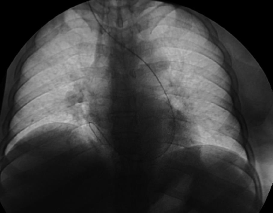
Figure 1 Digital venogram showing the curve of the metallic flexible guidewire from the left to the right atrium.
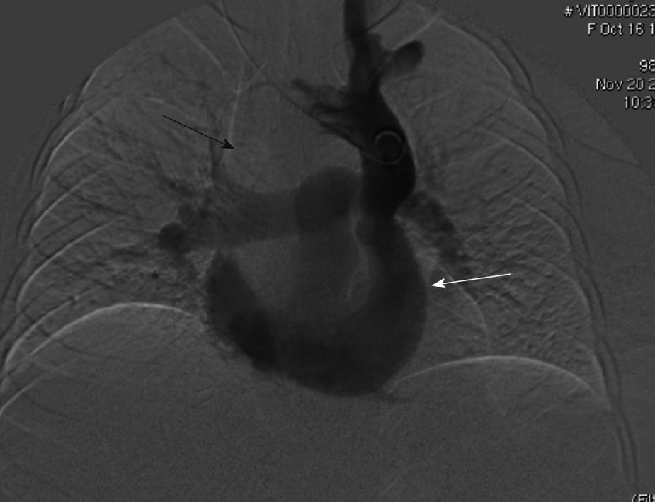
Figure 2 Digital venogram, performed with a 5F pigtail catheter, showing the absence of the right superior vena cava (RSVC) (black arrow) and presence of contrast dye in the left superior vena cava (LSVC) that is draining in the coronary sinus (white arrow).
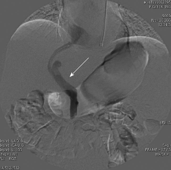
Figure 3 Digital portogram performed after transjugular intrahepatic portosystemic placement showing patency of the stent with good portal flow (white arrow).
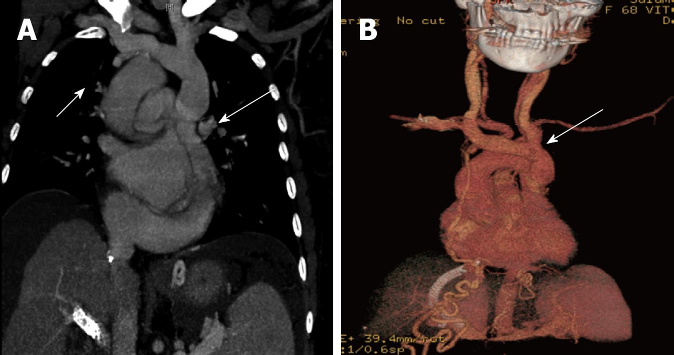
Figure 4 Maximum intensity projection reconstruction in coronal plane (A) and volume rendering reconstruction (B).
Absence of the right vena cava (short arrow) with the brachio-cephalic venous trunk, the left jugular vein and the left subclavian vein draining into the left superior vena cava (long arrows). Left SCV is draining into the coronary sinus.
- Citation: Petridis I, Miraglia R, Marrone G, Gruttadauria S, Luca A, Vizzini GB, Gridelli B. Transjugular intrahepatic portosystemic shunt with accidental diagnosis of persistence of the left superior vena cava. World J Gastroenterol 2010; 16(9): 1158-1160
- URL: https://www.wjgnet.com/1007-9327/full/v16/i9/1158.htm
- DOI: https://dx.doi.org/10.3748/wjg.v16.i9.1158


