
















©2010 Baishideng.
World J Gastroenterol. Mar 7, 2010; 16(9): 1158-1160
Published online Mar 7, 2010. doi: 10.3748/wjg.v16.i9.1158
Published online Mar 7, 2010. doi: 10.3748/wjg.v16.i9.1158
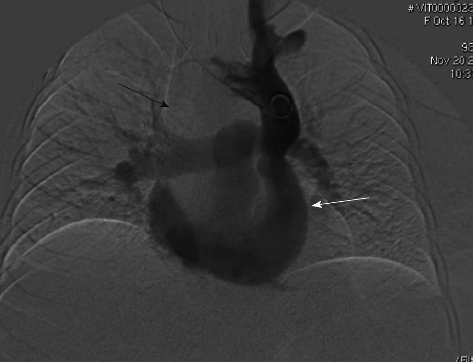
Figure 2 Digital venogram, performed with a 5F pigtail catheter, showing the absence of the right superior vena cava (RSVC) (black arrow) and presence of contrast dye in the left superior vena cava (LSVC) that is draining in the coronary sinus (white arrow).
- Citation: Petridis I, Miraglia R, Marrone G, Gruttadauria S, Luca A, Vizzini GB, Gridelli B. Transjugular intrahepatic portosystemic shunt with accidental diagnosis of persistence of the left superior vena cava. World J Gastroenterol 2010; 16(9): 1158-1160
- URL: https://www.wjgnet.com/1007-9327/full/v16/i9/1158.htm
- DOI: https://dx.doi.org/10.3748/wjg.v16.i9.1158