Copyright
©2010 Baishideng.
World J Gastroenterol. Feb 7, 2010; 16(5): 588-595
Published online Feb 7, 2010. doi: 10.3748/wjg.v16.i5.588
Published online Feb 7, 2010. doi: 10.3748/wjg.v16.i5.588
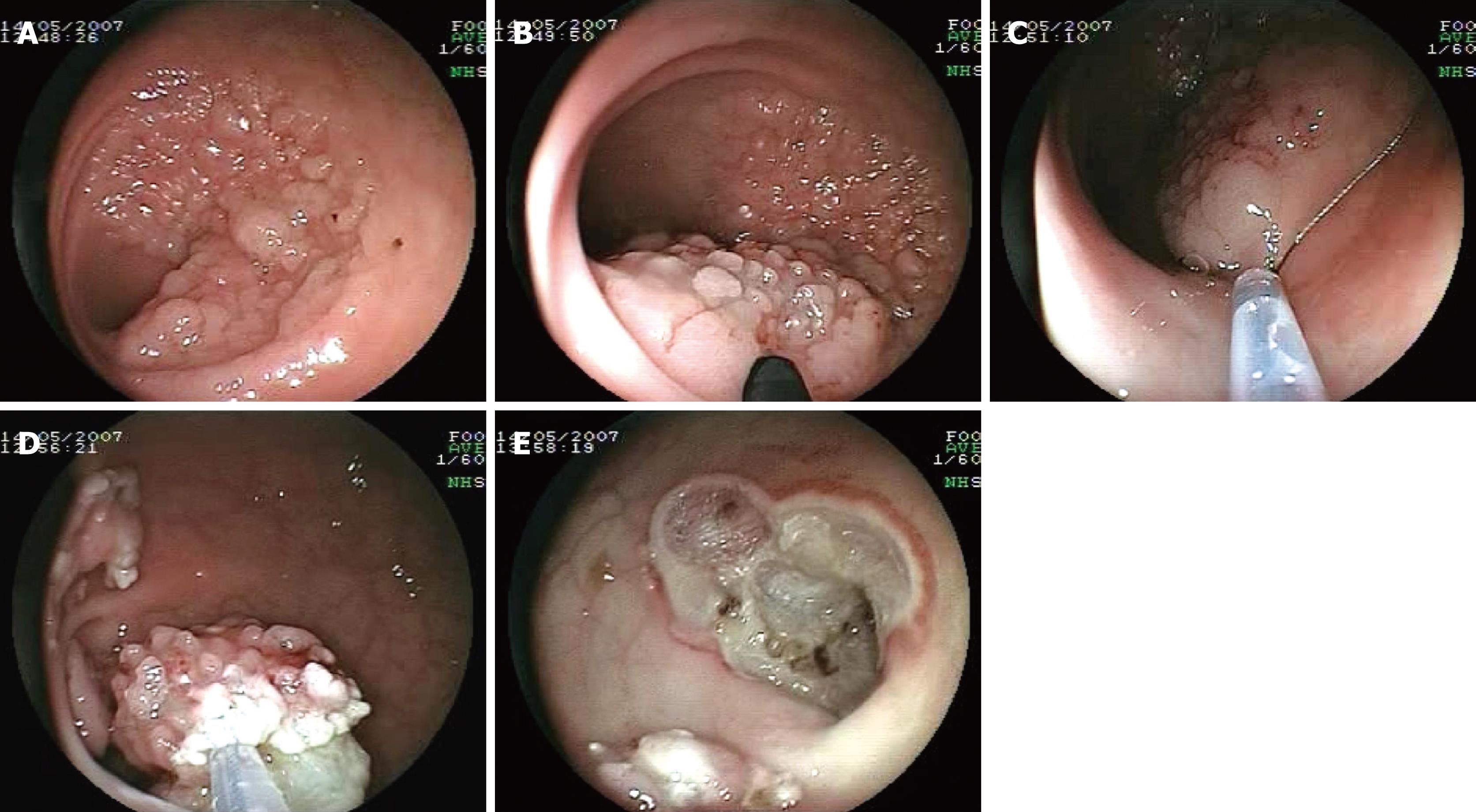
Figure 1 Endoscopic piecemeal mucosal resection image.
A: Large lateral spreading rectal tumor (adenoma with high grade dysplasia); B: Submucosal lifting of the tumor using saline with adrenaline 1/10 000; C: Capture of the lifted part of the tumor with a needle snare; D: Piecemeal resection of the rectal tumor; E: Final aspect at the end of the resection (procedure duration 70 min).
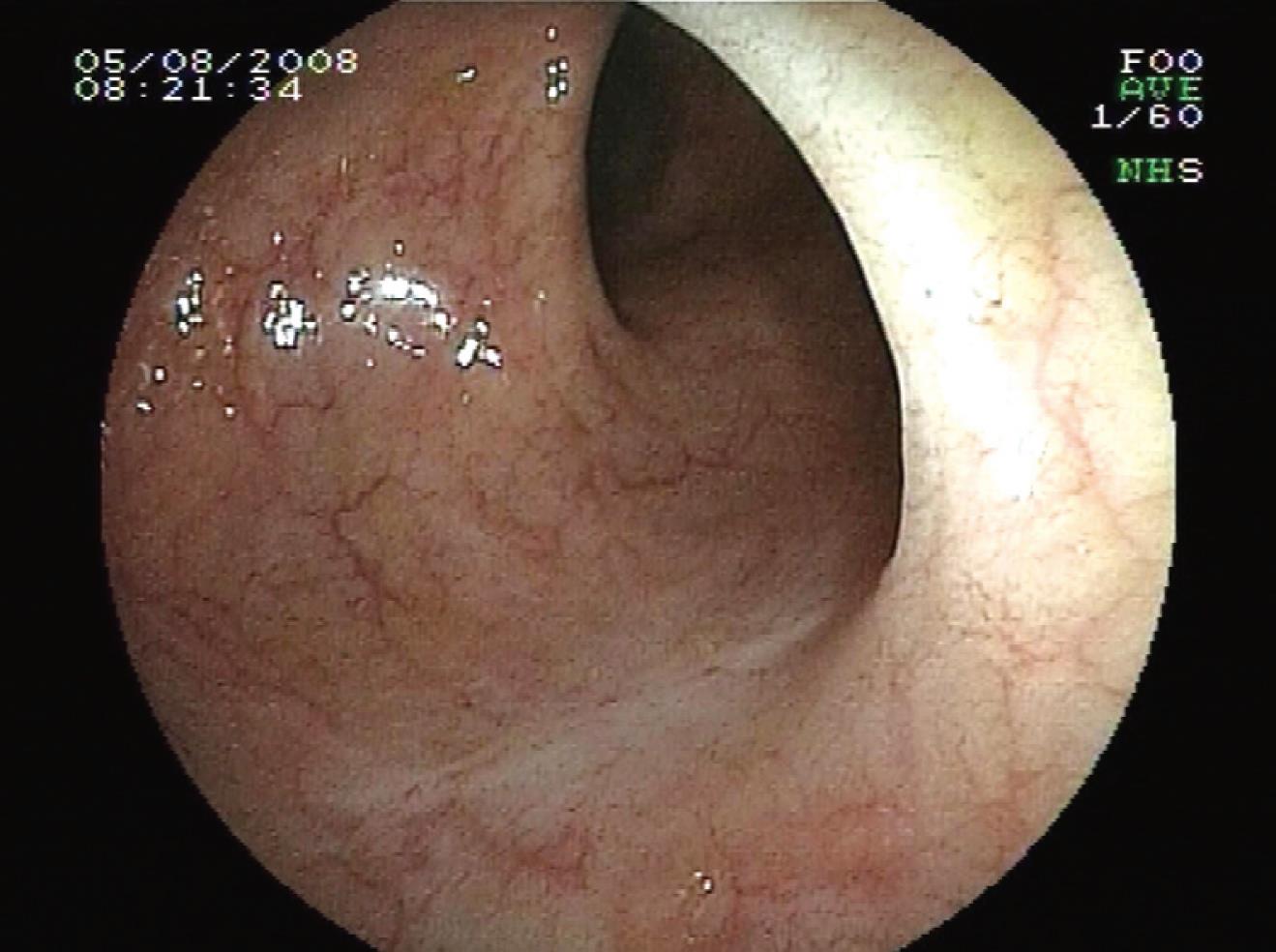
Figure 2 One year follow-up: a scar is visible without any sign of recurrence.
- Citation: Soune PA, Ménard C, Salah E, Desjeux A, Grimaud JC, Barthet M. Large endoscopic mucosal resection for colorectal tumors exceeding 4 cm. World J Gastroenterol 2010; 16(5): 588-595
- URL: https://www.wjgnet.com/1007-9327/full/v16/i5/588.htm
- DOI: https://dx.doi.org/10.3748/wjg.v16.i5.588


