Copyright
©2010 Baishideng.
World J Gastroenterol. Jan 28, 2010; 16(4): 518-521
Published online Jan 28, 2010. doi: 10.3748/wjg.v16.i4.518
Published online Jan 28, 2010. doi: 10.3748/wjg.v16.i4.518
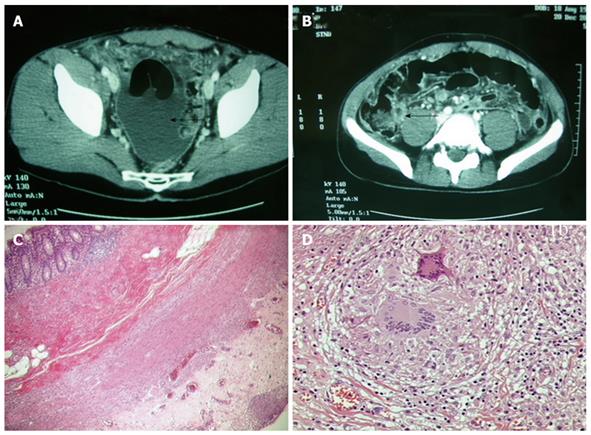
Figure 1 CT scan pre-operative images and histopathological images.
A: CT-scan image showing relevant amount of fluid in the Douglas; B: CT-scan image showing clear thickening of the appendicular wall (arrows) and “target” image as an evident sign of appendicitis; C, D: Histopathological aspects of the bioptic and resected samples.
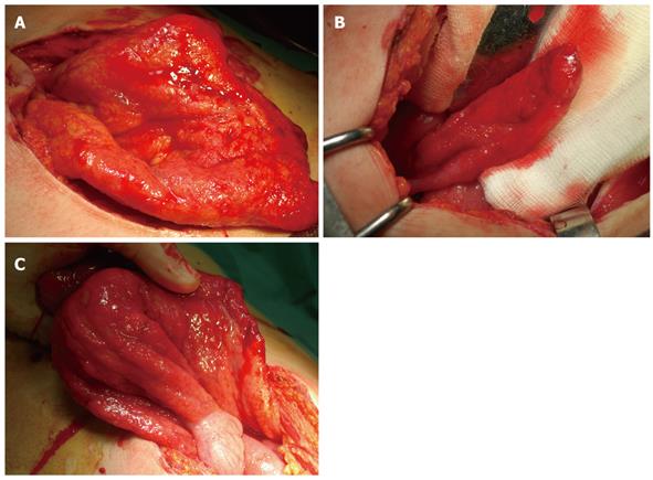
Figure 2 Specimen after laparotomy.
A: Diffuse military aspect, simulating a peritoneal carcinosis; B: Tenacious adhesions in the appendix-ileum-blind gut region; C: Diffuse inflammatory, hyperemic and edematous aspects of the intestine.
- Citation: Barbagallo F, Latteri S, Sofia M, Ricotta A, Castello G, Chisari A, Randazzo V, Greca GL. Appendicular tuberculosis: The resurgence of an old disease with difficult diagnosis. World J Gastroenterol 2010; 16(4): 518-521
- URL: https://www.wjgnet.com/1007-9327/full/v16/i4/518.htm
- DOI: https://dx.doi.org/10.3748/wjg.v16.i4.518


