Copyright
copy;2010 Baishideng Publishing Group Co.
World J Gastroenterol. Oct 21, 2010; 16(39): 4980-4985
Published online Oct 21, 2010. doi: 10.3748/wjg.v16.i39.4980
Published online Oct 21, 2010. doi: 10.3748/wjg.v16.i39.4980
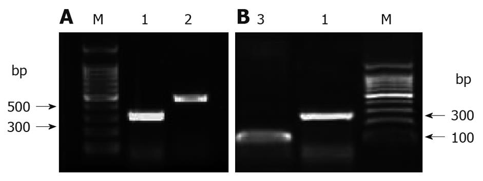
Figure 1 Basic expression of CABYR (A) and nuclear factor-κB (B) at mRNA level in 293T cells.
Total RNA was extracted from 293T cells with Trizol and reverse transcribed to 293T cDNA with the primer Oligo(dt). The target fragment was amplified by semi-quantitative polymerase chain reaction and analyzed by agarose electrophoresis (1%). CABYR and nuclear factor (NF)-κB were detectable in 293T cells, indicating that 293T cells can be used to identify the efficient silence fragment for CABYR and study the relation between CABYR and NF-κB. 1: β-actin; 2: CABYR; 3: NF-κB; M: Marker.
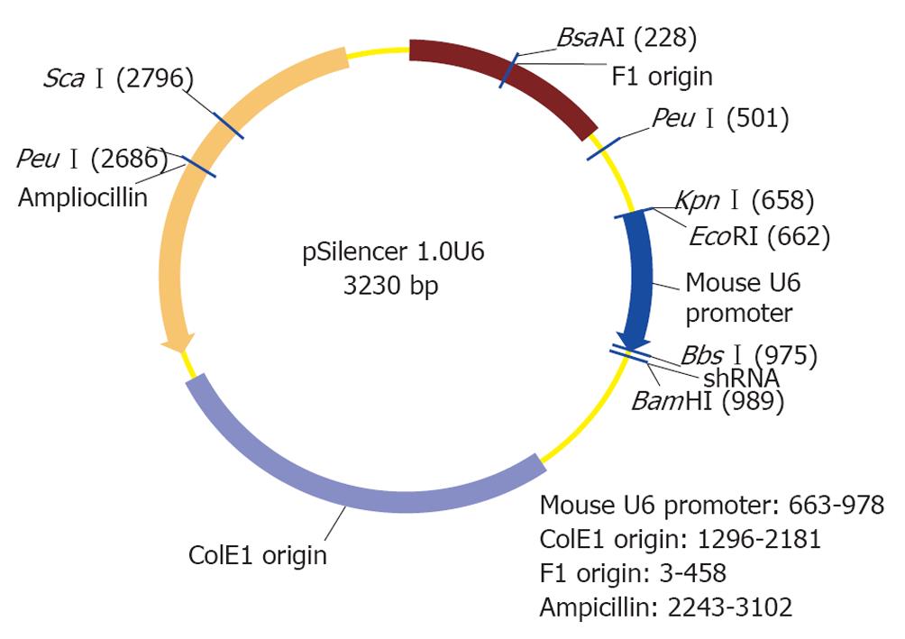
Figure 2 Construction of CABYR shRNA eucaryon expression vector by inserting the target gene fragment into pSlience1.
0 (3.23 kb) between BbsI and BamHI.
Figure 3 Construction of CABYR shRNA eucaryon expression vector by inserting the target fragment we constructed into the RNAi plasmid.
1:vacant vector; 2: Cabyrmid 1 vector; 3: Cabyrmid 2 vector; 4: Cabyrmid 3 vector; M1: Marker 1; M2: Marker 2.
Figure 4 Construction of CABYR shRNA eucaryon expression vector by locating shRNA in plasmid.
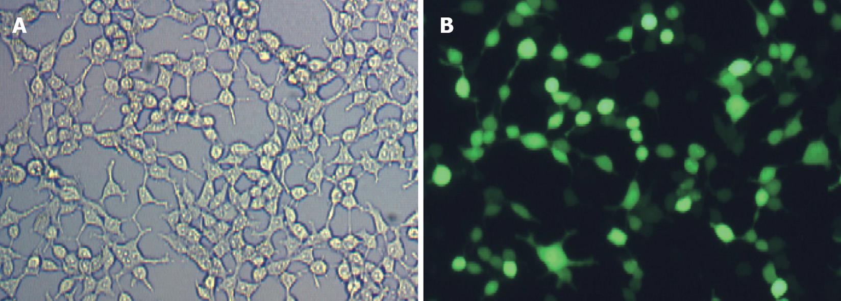
Figure 5 CABYR shRNA expression in reference group (A) and CABYR mRNA expression in other groups (B).
A: HE stain, × 100; B: Blue fluorescent, ×100.
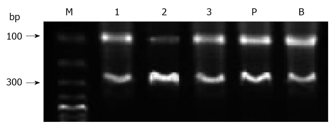
Figure 6 CABYR mRNA expressions in different groups after transfection.
M: Marker; 1: Cabyrmid 1; 2: Cabyrmid 2; 3: Cabyrmid 3; P: Vacant vector; B: Blank.
- Citation: Shi LX, He YM, Fang L, Meng HB, Zheng LJ. CABYR RNAi plasmid construction and NF-κB signal transduction pathway. World J Gastroenterol 2010; 16(39): 4980-4985
- URL: https://www.wjgnet.com/1007-9327/full/v16/i39/4980.htm
- DOI: https://dx.doi.org/10.3748/wjg.v16.i39.4980


