Copyright
copy;2010 Baishideng Publishing Group Co.
World J Gastroenterol. Sep 28, 2010; 16(36): 4621-4624
Published online Sep 28, 2010. doi: 10.3748/wjg.v16.i36.4621
Published online Sep 28, 2010. doi: 10.3748/wjg.v16.i36.4621
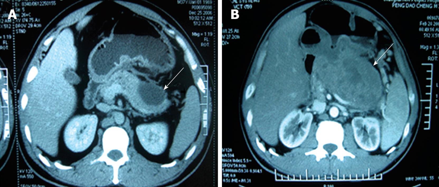
Figure 1 Computed tomography showing a pseudocystic tumor (arrows) as a benign cyst (A) and a bigger pseudocystic tumor poorly demarcated from the surrounding tissue (B).
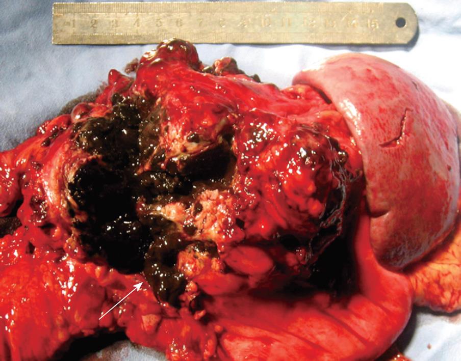
Figure 2 Specimen showing a metastatic melanoma as a black-brown mass (arrow) in pancreatic tail.
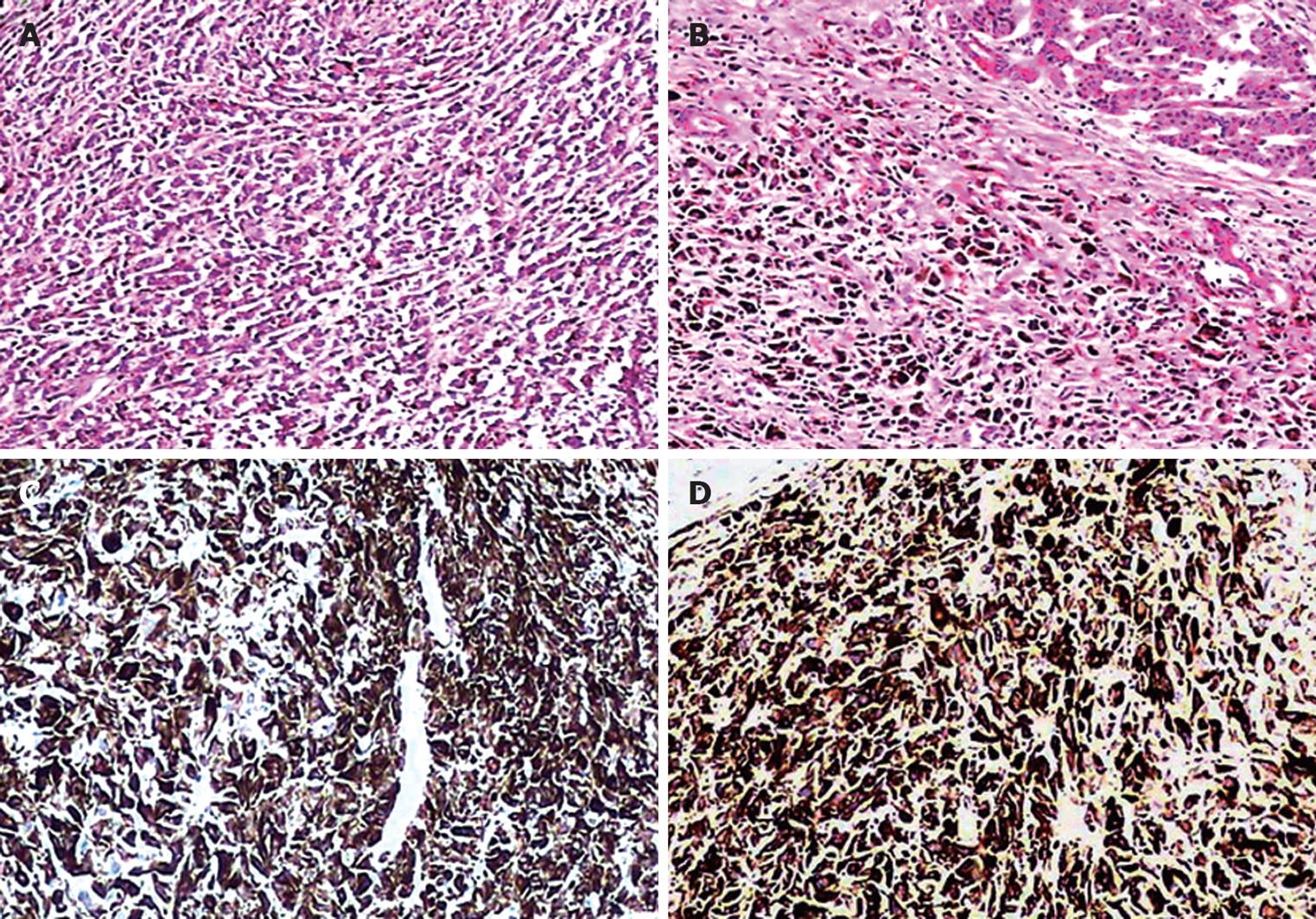
Figure 3 Microscopy showing epithelioid or polygon tumor cells infiltrating the pancreatic tail (A), dark brown granular intracytoplasmic pigmentation in most tumor cells (B), positive HMB-45 (C) and S-100 protein (D).
Original magnification × 200. A and B: HE staining; C and D: Immunostaining.
- Citation: He MX, Song B, Jiang H, Hu XG, Zhang YJ, Zheng JM. Complete resection of isolated pancreatic metastatic melanoma: A case report and review of the literature. World J Gastroenterol 2010; 16(36): 4621-4624
- URL: https://www.wjgnet.com/1007-9327/full/v16/i36/4621.htm
- DOI: https://dx.doi.org/10.3748/wjg.v16.i36.4621


