Copyright
copy;2010 Baishideng Publishing Group Co.
World J Gastroenterol. Sep 28, 2010; 16(36): 4616-4620
Published online Sep 28, 2010. doi: 10.3748/wjg.v16.i36.4616
Published online Sep 28, 2010. doi: 10.3748/wjg.v16.i36.4616
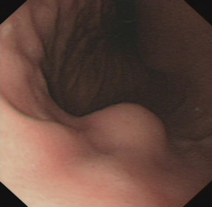
Figure 1 Upper gastrointestinal endoscopy showing a well-circumscribed elevated mass lesion measuring 1.
5 cm × 1.5 cm with normal overlying mucosa in anterior wall of gastric corpus.
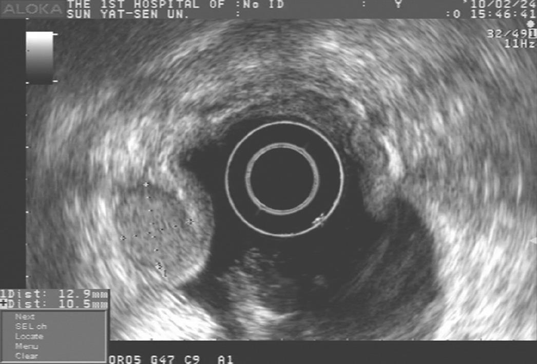
Figure 2 Endoscopic ultrasonography showing a 1.
5 cm × 1.2 cm sharply demarcated homogeneous hypoechoic mass in the third and fourth sonographic layers of gastric wall.
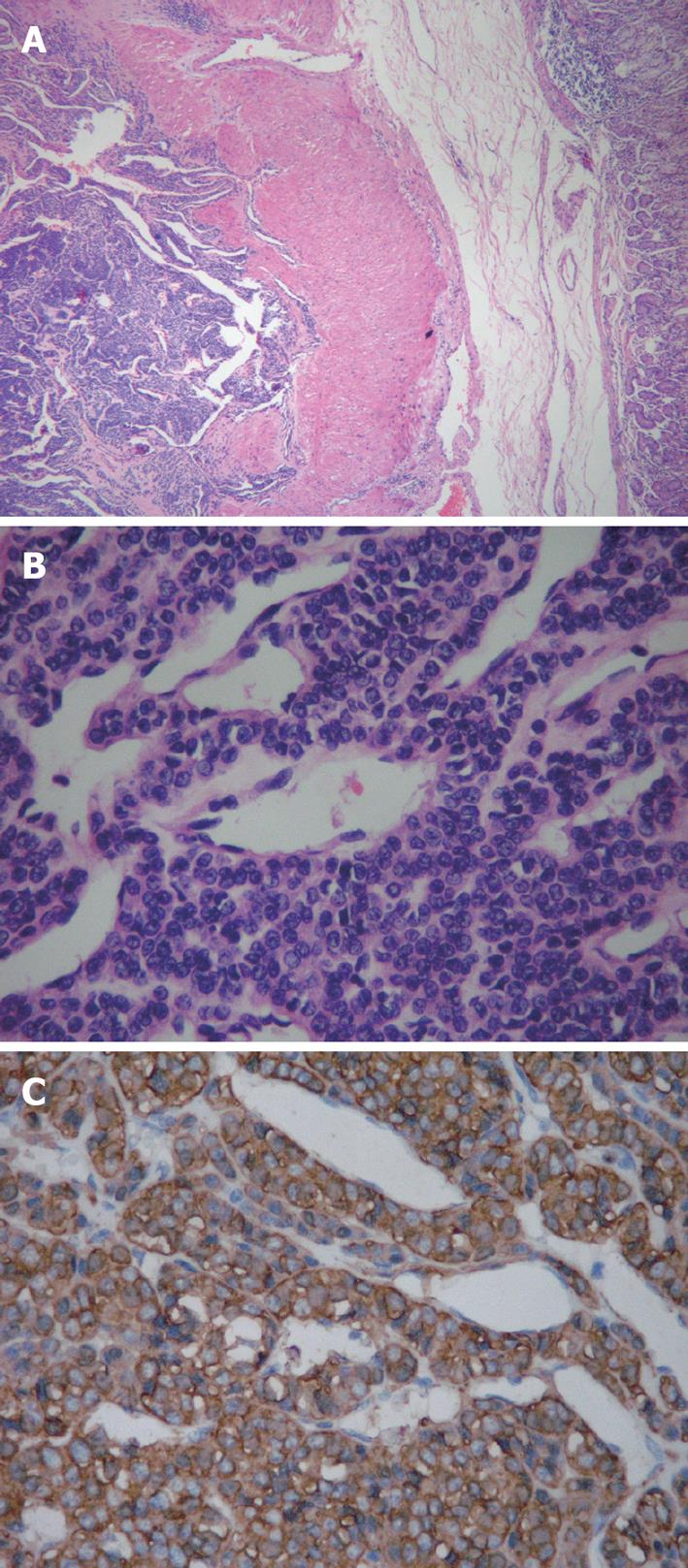
Figure 3 Gastric glomus tumor.
A: Glomus tumor located in the muscularis of stomach (hematoxylin-eosin × 40); B: Clusters of uniform, round tumor cells around dilated blood vessels (hematoxylin-eosin × 200); C: Tumor cells positive for smooth muscle actin (immunohistochemistry staining × 200).
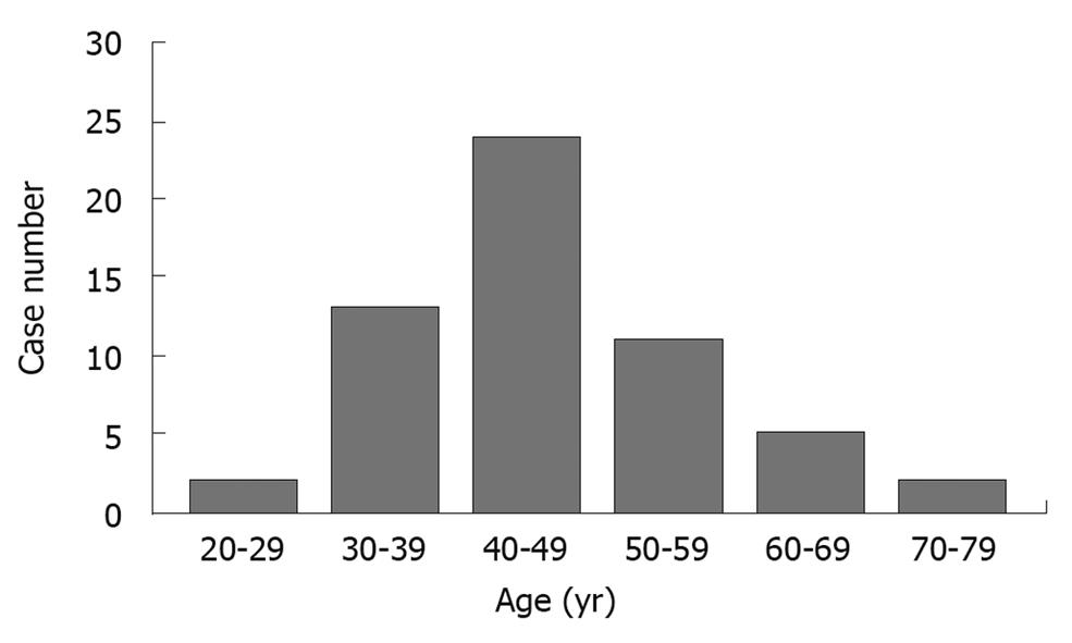
Figure 4 Distribution of age in 57 cases of gastric glomus tumor.
The age distribution of 57 cases of gastric glomus tumors
- Citation: Fang HQ, Yang J, Zhang FF, Cui Y, Han AJ. Clinicopathological features of gastric glomus tumor. World J Gastroenterol 2010; 16(36): 4616-4620
- URL: https://www.wjgnet.com/1007-9327/full/v16/i36/4616.htm
- DOI: https://dx.doi.org/10.3748/wjg.v16.i36.4616


