Copyright
©2010 Baishideng Publishing Group Co.
World J Gastroenterol. Sep 14, 2010; 16(34): 4367-4370
Published online Sep 14, 2010. doi: 10.3748/wjg.v16.i34.4367
Published online Sep 14, 2010. doi: 10.3748/wjg.v16.i34.4367
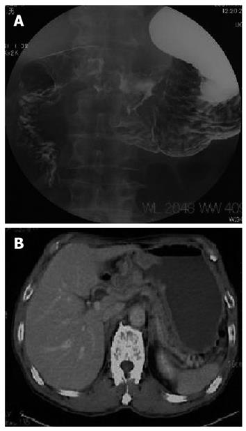
Figure 1 Gastrografin meal study revealing a tumor with ulceration at the gastric antrum, causing gastric outlet obstruction (A), and abdominal computed tomography scan showing remarkable thickness of gastric wall with no evidence of direct invasion to the pancreas head, and lymph node metastasis or distant metastasis (B).
Figure 2 Laparoscopy-assisted partitioning gastrojejunostomy conducted in a Roux-en Y fashion in an antecolic manner (A), cutting the tunnel as a single procedure in reconstruction of the second surgery after chemotherapy (B), and the completely excised tumor with distal gastrectomy and D2 lymph node dissection (C).
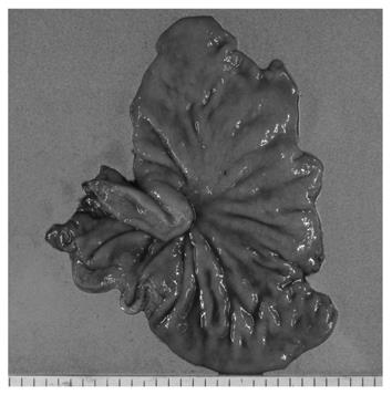
Figure 3 Resected specimen showing a grossly shallow depressed lesion as only fibrosis at the antrum with live cancer cells found in the fibrotic tissue throughout the whole gastric wall at histopathological examination.
- Citation: Okumura Y, Ohashi M, Nunobe S, Iwanaga T, Kanda T, Iwasaki Y. Gastrojejunostomy followed by induction chemotherapy for incurable gastric cancer with outlet obstruction. World J Gastroenterol 2010; 16(34): 4367-4370
- URL: https://www.wjgnet.com/1007-9327/full/v16/i34/4367.htm
- DOI: https://dx.doi.org/10.3748/wjg.v16.i34.4367


