Copyright
©2010 Baishideng.
World J Gastroenterol. Jan 21, 2010; 16(3): 365-371
Published online Jan 21, 2010. doi: 10.3748/wjg.v16.i3.365
Published online Jan 21, 2010. doi: 10.3748/wjg.v16.i3.365
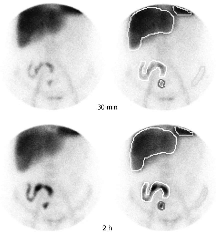
Figure 1 Regions of interest (ROIs).
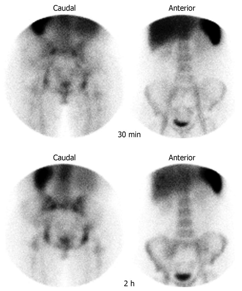
Figure 2 Images of a healthy volunteer (control) in the anterior and caudal projections at 30 min and at 2 h after injection of radiolabeled leukocytes.
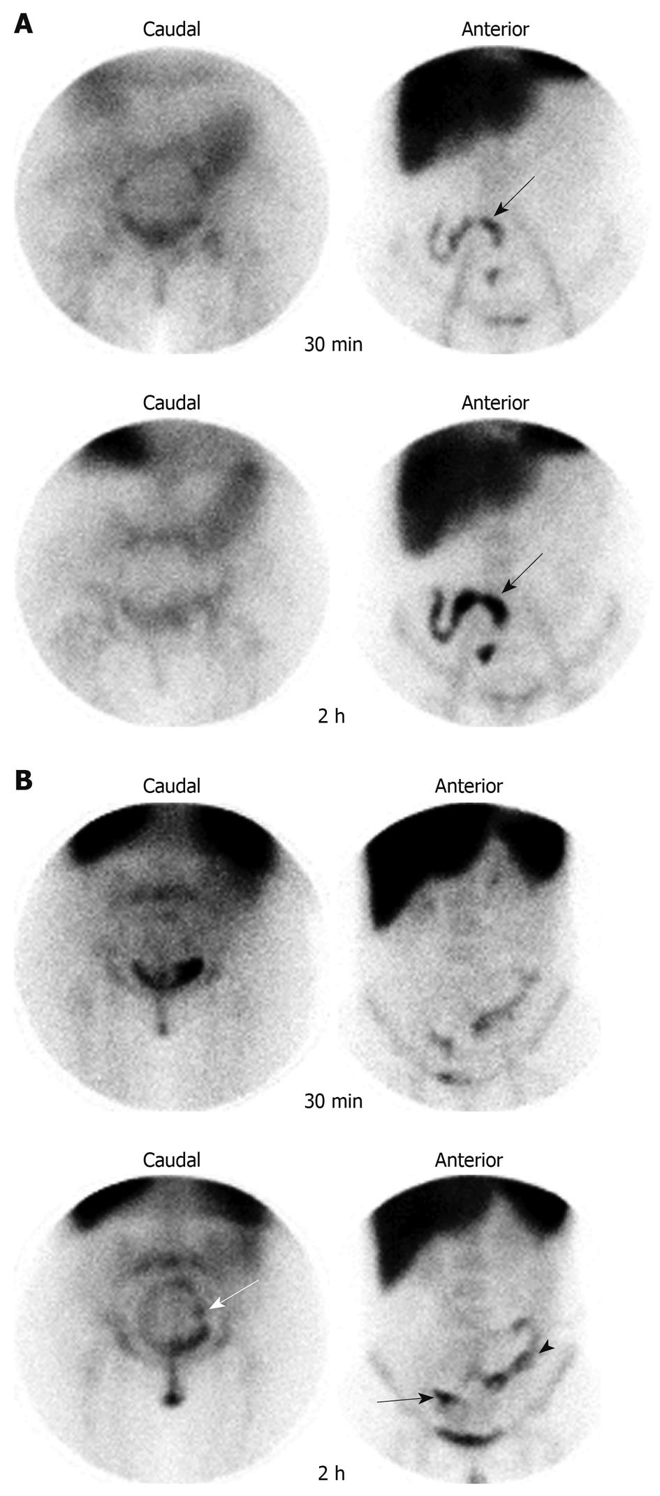
Figure 3 Images obtained 30 min and 2 h post-injection of 99mTc-HMPAO-labeled leukocytes in the caudal and anterior views.
A: The arrows indicate the region of the intestine affected by the inflammation (L.M.T., female, 54 years); B: The arrows indicate 99mTc-HMPAO-labeled leukocyte uptake in the terminal ileum, descending colon and rectum-sigmoid (L.D., female, 24 years). The black arrows indicate terminal ileum; the arrowhead indicates descending colon; the white arrow indicates rectum-sigmoid.
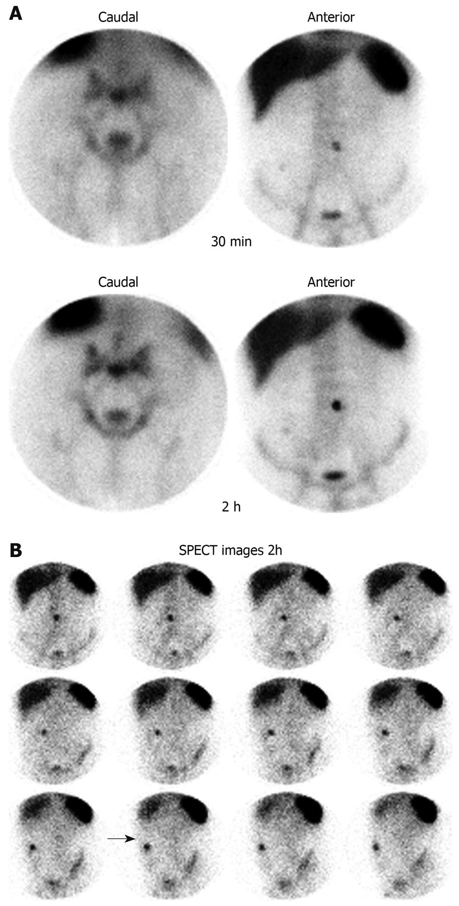
Figure 4 99mTc-HMPAO granulocyte scintigraphy of patient J.
A.N., male, 44 years. A: Images obtained at 30 min and at 2 h after injection of the radiolabeled leukocytes, in the caudal and anterior projections; B: SPECT images 2 h after injection of the radiolabeled leukocytes. The arrow indicates the fistula region of the patient’s abdomen.
- Citation: Mota LG, Coelho LG, Simal CJ, Ferrari ML, Toledo C, Martin-Comin J, Diniz SO, Cardoso VN. Leukocyte-technetium-99m uptake in Crohn’s disease: Does it show subclinical disease? World J Gastroenterol 2010; 16(3): 365-371
- URL: https://www.wjgnet.com/1007-9327/full/v16/i3/365.htm
- DOI: https://dx.doi.org/10.3748/wjg.v16.i3.365


