Copyright
©2010 Baishideng.
World J Gastroenterol. Jul 28, 2010; 16(28): 3546-3552
Published online Jul 28, 2010. doi: 10.3748/wjg.v16.i28.3546
Published online Jul 28, 2010. doi: 10.3748/wjg.v16.i28.3546
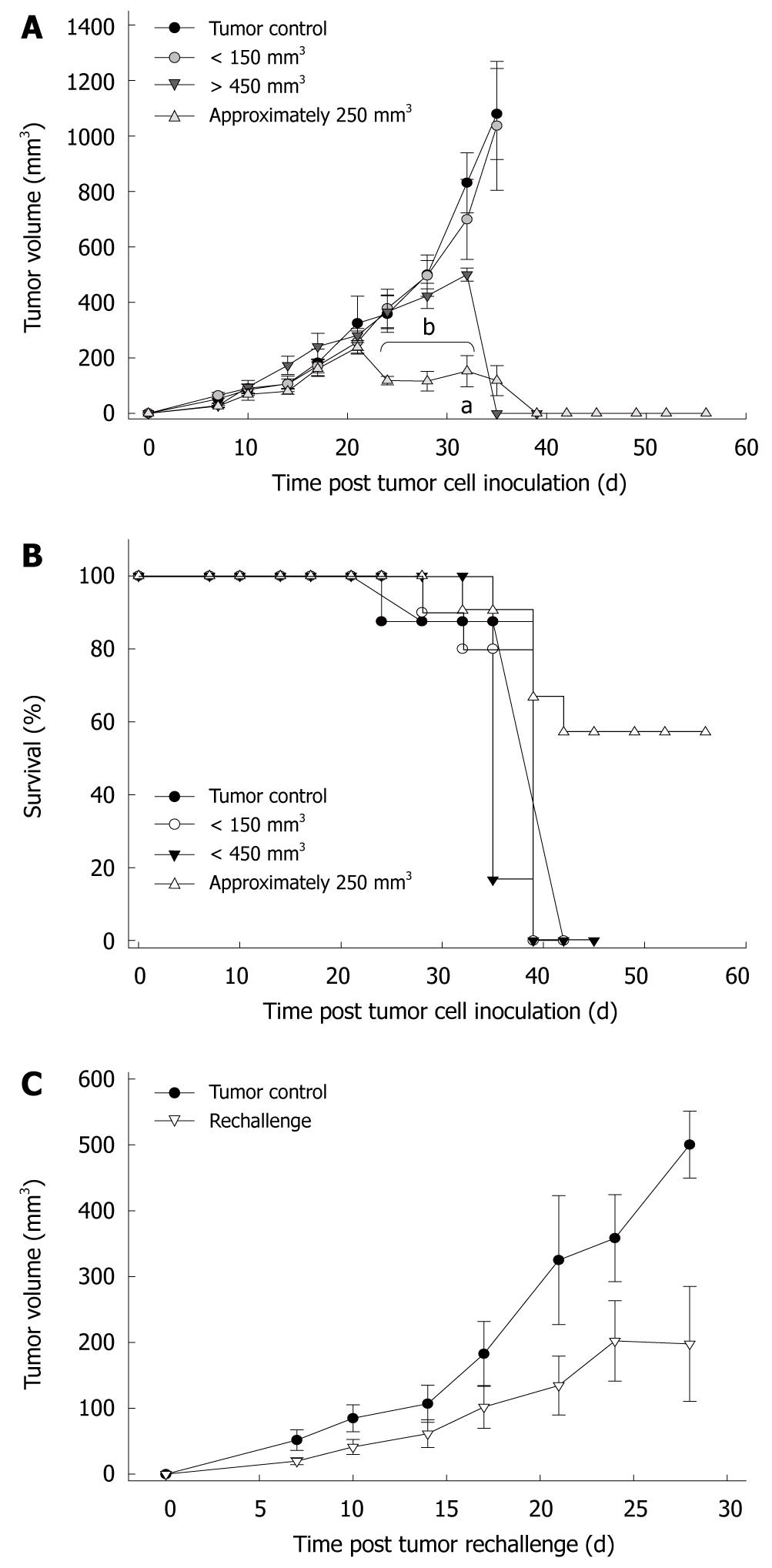
Figure 1 Data of in vivo-analyses of s.
c. Panc02 tumors treated with Clostridium novyi-NT spores. A: Growth kinetics of s.c. Panc02 tumors; B: Survival curve of tumor-carrying mice after i.v. injection of Clostridium novyi-NT spores. For evaluation of the optimal therapeutic dose, different groups of tumor carrying mice were employed. These included tumor sizes below 150 mm3 (n = 10), of about 250 mm3 (n = 21) and larger than 450 mm3 (n = 6). Experiments identified a tumor-size dependent toxicity and response rate. Small tumors were completely unaffected and remained comparable to controls. In contrast, larger tumors responded with substantial necrosis followed by shrinkage and subsequent complete regression; C: Growth kinetics of tumors after rechallenge. Successfully treated animals (n = 5) received a second tumorigenic dose of Panc02 cells at day 28 post therapy. Naïve animals were used as controls. As can be depicted from the graph, bacteriolytic therapy mediated partial protection from re-exposure to tumor cells. Values are given as the mean tumor volume (mm3) ± SE; aP < 0.05 vs tumor control; bP < 0.001 vs tumor control; Mann-Whitney U-test.
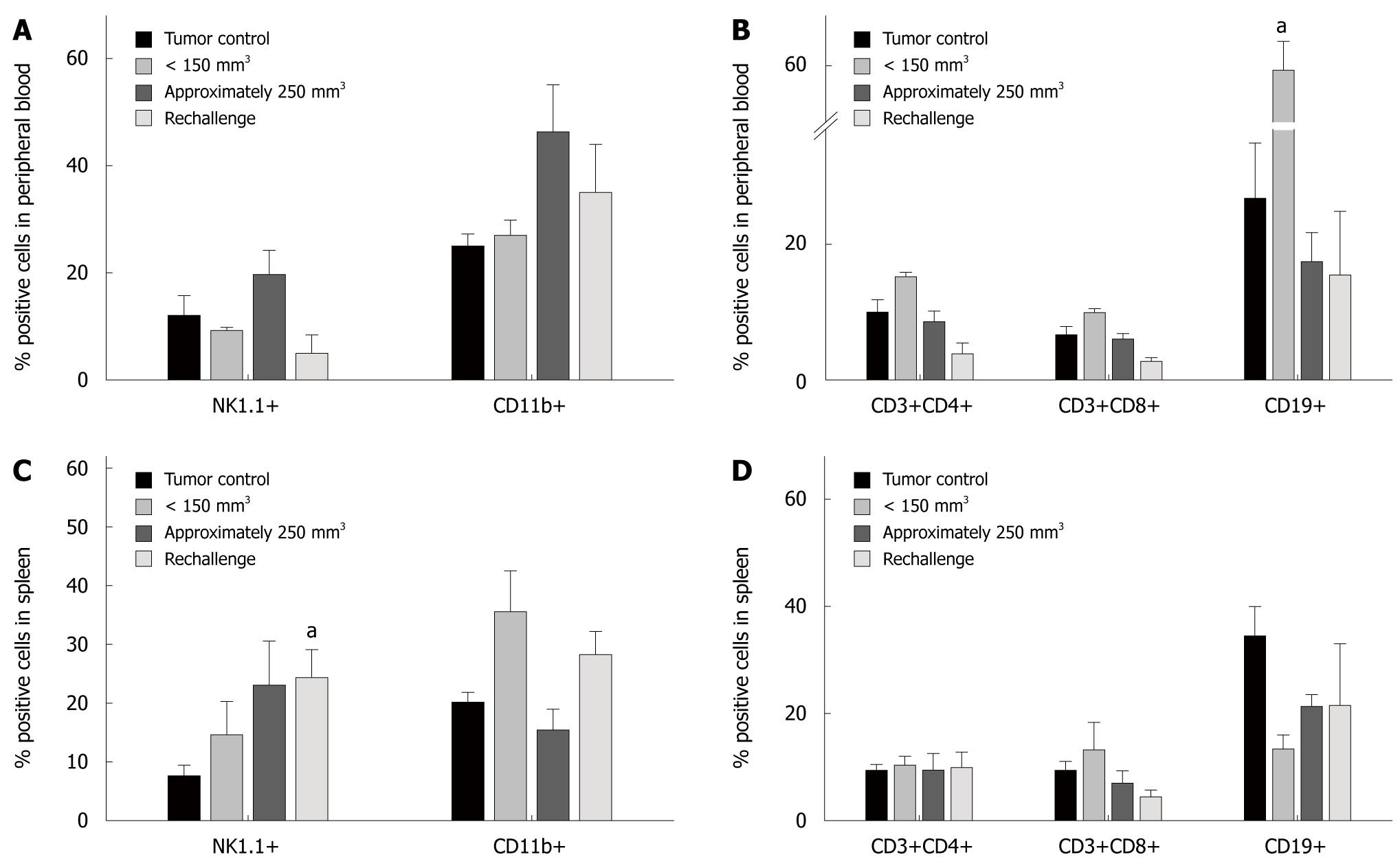
Figure 2 Flow cytometric analyses of leukocytes from peripheral blood (A, B) and spleens (C, D) in tumor control animals, of Clostridium novyi-NT treated animals (< 150 mm3, approximately 250 mm3) and after rechallenge.
Bacteriolytic therapy predominantly activated the innate arm of the immune system. Values are given as mean ± SE. aP < 0.05 vs tumor control; Mann-Whitney U-test.
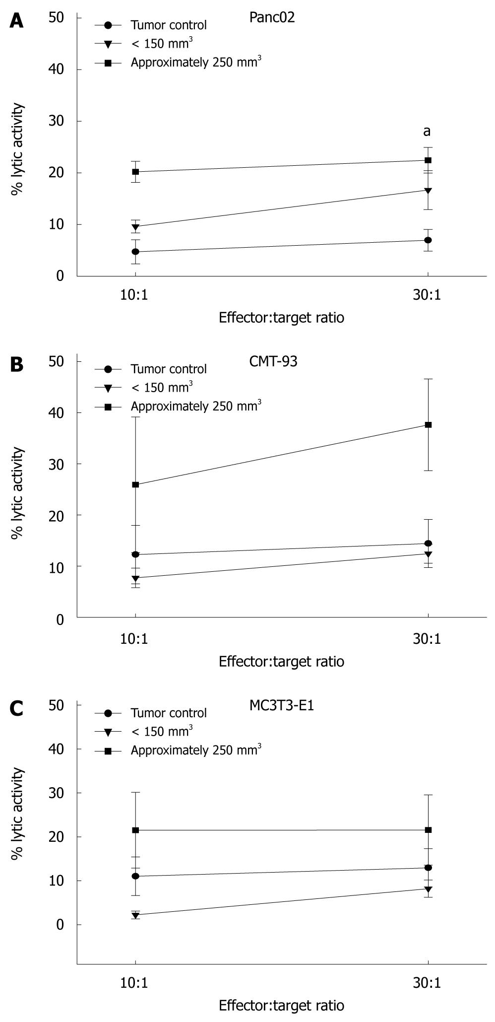
Figure 3 Quantitative analysis of cytotoxicity using flow cytometric carboxylfluorescein diacetate/propidium iodide staining.
Lymphocytes were isolated from spleens and co-cultured with targets for five hours at E:T cell ratios of 10:1 and 30:1. Lymphocytes from successfully treated animals showed lytic activity against (A, B) syngeneic tumor cells with highest reactivity against CMT-93 cells at an E:T ratio of 30:1. However, cytotoxicity was found to be non-tumor specific, as reactivity was also observed against (C) non-malignant fibroblasts MC3T3-E1. Values are given as mean ± SE; aP < 0.05 vs tumor control; t-test.
- Citation: Maletzki C, Gock M, Klier U, Klar E, Linnebacher M. Bacteriolytic therapy of experimental pancreatic carcinoma. World J Gastroenterol 2010; 16(28): 3546-3552
- URL: https://www.wjgnet.com/1007-9327/full/v16/i28/3546.htm
- DOI: https://dx.doi.org/10.3748/wjg.v16.i28.3546


