Copyright
©2010 Baishideng.
World J Gastroenterol. Jun 28, 2010; 16(24): 3083-3086
Published online Jun 28, 2010. doi: 10.3748/wjg.v16.i24.3083
Published online Jun 28, 2010. doi: 10.3748/wjg.v16.i24.3083
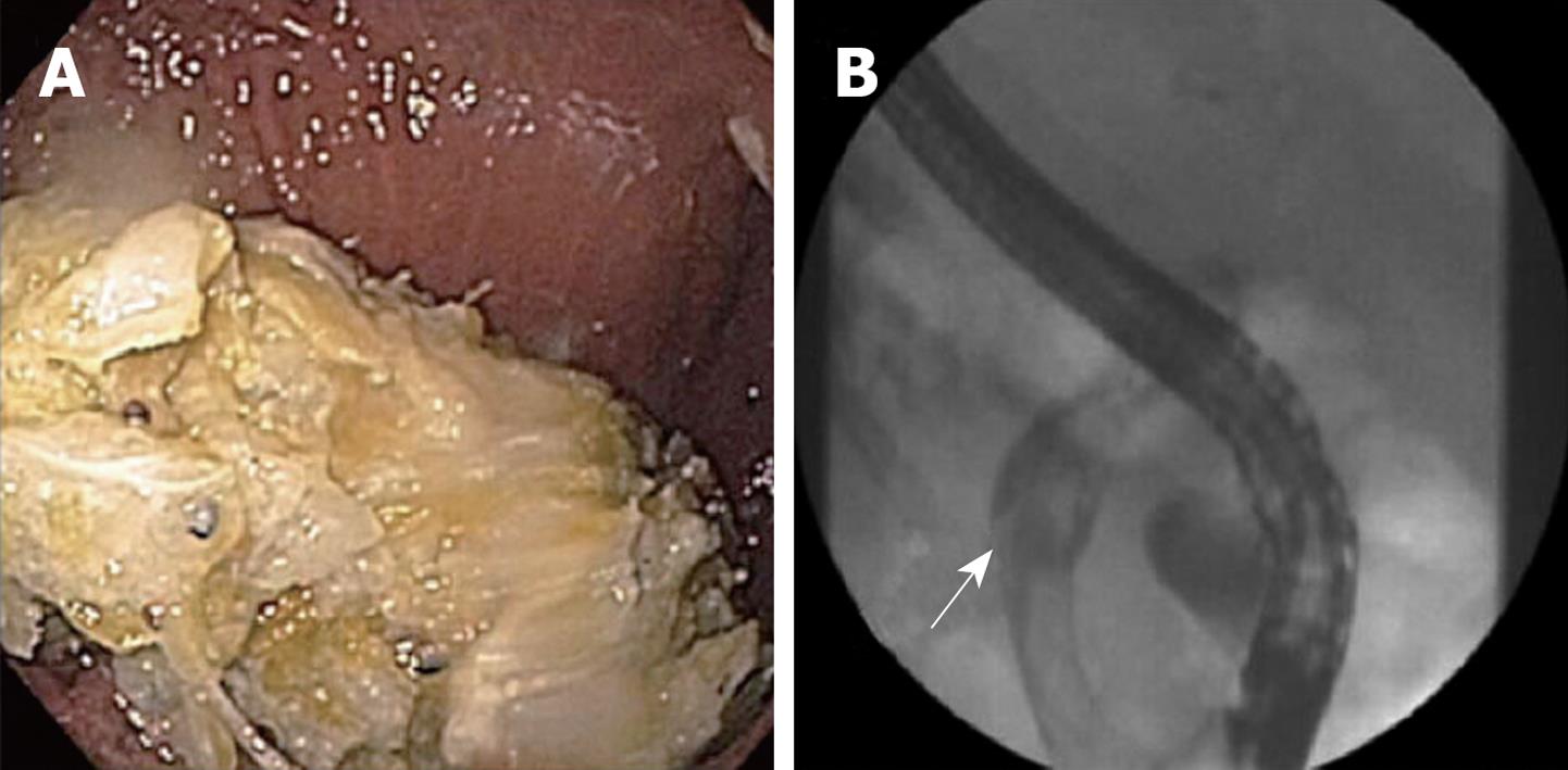
Figure 1 Endoscopic and cholangiographic views of biliary obstruction in patient 1.
A: Biliary/fungal debris occluding metal biliary stent; B: “Fungal balls” (arrow) obstructing the bile ducts by cholangiography.
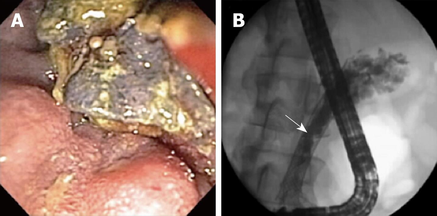
Figure 2 Endoscopic and cholangiographic views of biliary obstruction in patient 2.
A: “Fungal ball” visible in the common bile duct; B: Cholangiogram showing fungal debris (arrow) around the metal biliary stent.
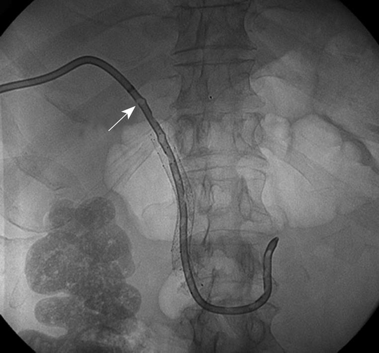
Figure 3 Cholangiographic view of fungal balls (ex.
arrow) within the percutaneous transhepatic biliary drainage catheter in case 3.
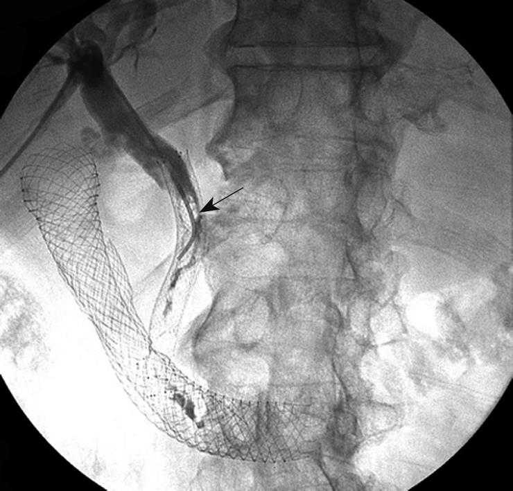
Figure 4 Cholangiographic view of fungal debris (arrow) within a metal biliary stent in case 4.
- Citation: Story B, Gluck M. Obstructing fungal cholangitis complicating metal biliary stent placement in pancreatic cancer. World J Gastroenterol 2010; 16(24): 3083-3086
- URL: https://www.wjgnet.com/1007-9327/full/v16/i24/3083.htm
- DOI: https://dx.doi.org/10.3748/wjg.v16.i24.3083


