Copyright
©2010 Baishideng.
World J Gastroenterol. Jun 7, 2010; 16(21): 2698-2701
Published online Jun 7, 2010. doi: 10.3748/wjg.v16.i21.2698
Published online Jun 7, 2010. doi: 10.3748/wjg.v16.i21.2698
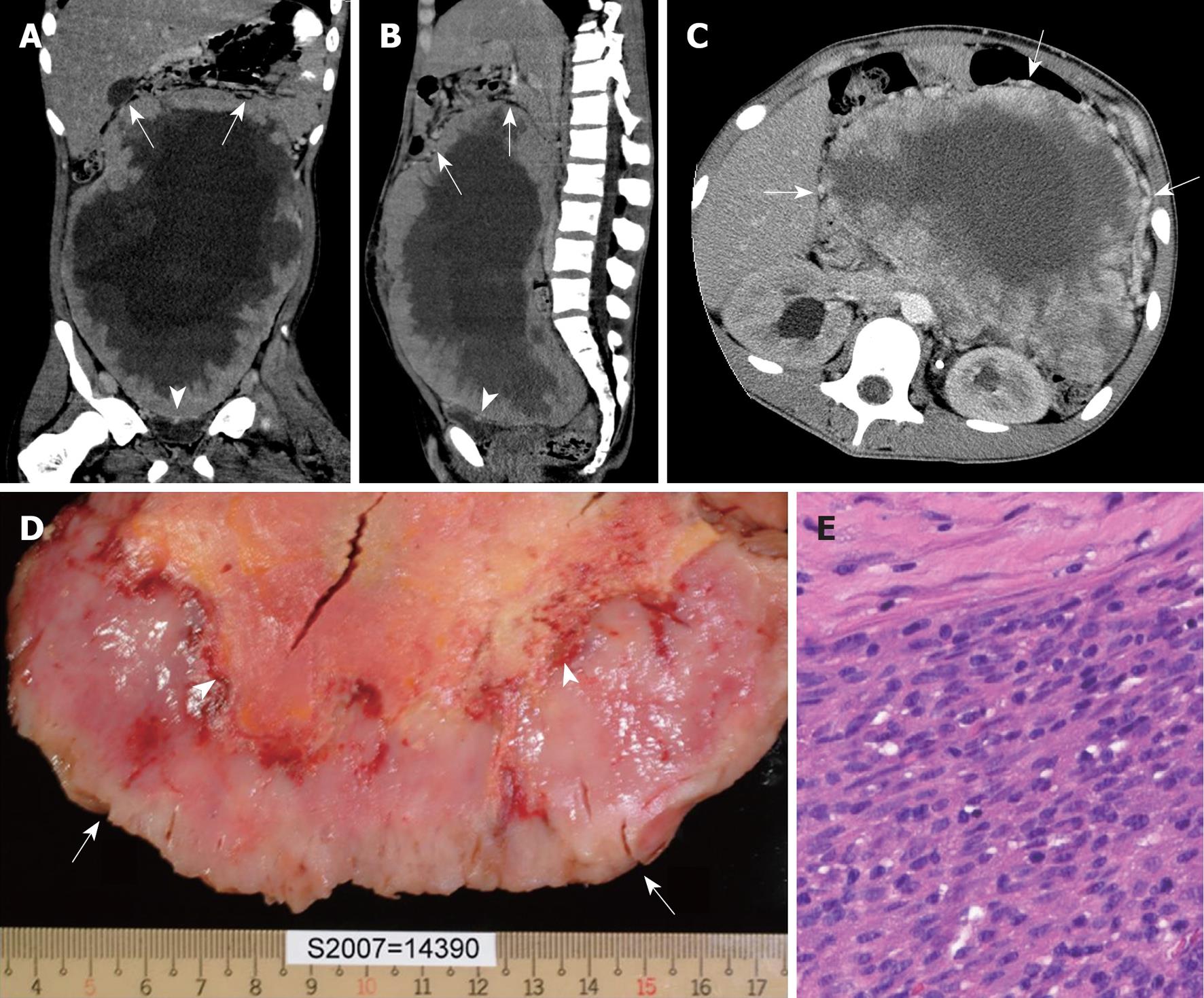
Figure 1 Malignant inflammatory myofibroblastic tumor in a 14-year-old with a history of abdominal pain and body weight loss.
A, B: Coronal (A) and sagittal (B) computed tomography (CT) images reveal a huge pelvi-abdominal mass with an uneven peripheral solid component and massive central necrosis, with upward displacement of the bowel loops (arrows) and downward compression on the urinary bladder (arrowheads), highly suggestive of an extraperitoneal tumor originating from the perivesical space; C: Axial CT image at the level of the kidney clearly demonstrates prominent vessels around the tumor (arrows); D: The cut-surface of the gross specimen reveals an uneven peripheral solid component (arrows) and massive central necrosis (arrowheads); E: Photomicrograph shows proliferation of spindle tumor cells in the peripheral solid part (HE stain, original magnification × 200).
- Citation: Lu CH, Huang HY, Chen HK, Chuang JH, Ng SH, Ko SF. Huge pelvi-abdominal malignant inflammatory myofibroblastic tumor with rapid recurrence in a 14-year-old boy. World J Gastroenterol 2010; 16(21): 2698-2701
- URL: https://www.wjgnet.com/1007-9327/full/v16/i21/2698.htm
- DOI: https://dx.doi.org/10.3748/wjg.v16.i21.2698


