Copyright
©2010 Baishideng.
World J Gastroenterol. May 21, 2010; 16(19): 2440-2442
Published online May 21, 2010. doi: 10.3748/wjg.v16.i19.2440
Published online May 21, 2010. doi: 10.3748/wjg.v16.i19.2440
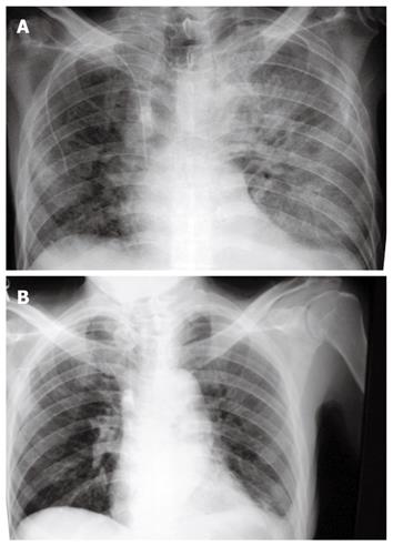
Figure 1 Chest X-ray.
A: Chest X-ray taken at time of admission showing Acute respiratory distress syndrome (ARDS); B: Chest X-ray after surgical colectomy showing a significant improvement of ARDS.
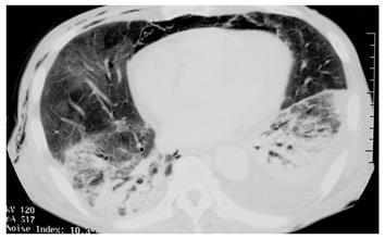
Figure 2 Chest computed tomography (CT) scan image taken before surgical treatment showing compatibility with ARDS.
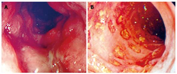
Figure 3 Colonoscopic images showing deep ulcer with severe mucosal edema (A) and multiple discrete ulcers (B).
- Citation: Sagara S, Horie Y, Anezaki Y, Miyazawa H, Iizuka M. Acute respiratory distress syndrome associated with severe ulcerative colitis. World J Gastroenterol 2010; 16(19): 2440-2442
- URL: https://www.wjgnet.com/1007-9327/full/v16/i19/2440.htm
- DOI: https://dx.doi.org/10.3748/wjg.v16.i19.2440


