Copyright
©2010 Baishideng.
World J Gastroenterol. May 21, 2010; 16(19): 2341-2347
Published online May 21, 2010. doi: 10.3748/wjg.v16.i19.2341
Published online May 21, 2010. doi: 10.3748/wjg.v16.i19.2341
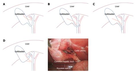
Figure 1 Anatomy of laparoscopic cholecystectomy (LC) (blue curves stand for bile duct and gallbladder and red curves for arteries).
A: Illustration of a variant cystic artery originating from the right hepatic artery; B: Cystic duct connected to the left wall of hepatic duct via an anterior approach; C: Cystic duct parallel with common hepatic duct; D: Cystic duct connected to the right hepatic duct; E: Calot’s triangle and Rouviere sulcus in LC.
Figure 2 Illustration of inguinal hernia structures.
A: Direct hernia was located medially to the inferior epigastric artery, an indirect hernia was located laterally to the inferior epigastric artery, and a femoral hernia was located at the inferior iliopubic tract; B: The three regions, where staples or sutures should not be placed: the triangle of doom, triangle of pain, and corona mortis; C: Live procedure after the peritoneum was absolutely dissected and removed from the abdominal wall.
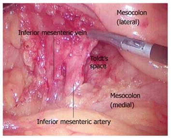
Figure 3 The root of inferior mesenteric artery.
The inferior mesenteric artery and vein were identified when the superior sigmoid colon mesentery was lifted to the left side. Toldt’s space was found in the included angle of the inferior mesenteric artery and abdominal aorta.
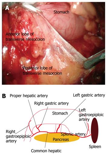
Figure 4 Anatomy of LRG.
A: Dissection in the first surgical plane, i.e. the space between anterior and posterior lobes of the transverse mesocolon; B: The main vessels in the space of the posterior layer of dorsal mesogastrium and pancreas when the paries posterior gastricus was turned over to the head side.
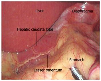
Figure 5 The lesser omentum above the hepatic caudate lobe.
- Citation: Li LJ, Zheng XM, Jiang DZ, Zhang W, Shen HL, Shan CX, Liu S, Qiu M. Progress in laparoscopic anatomy research: A review of the Chinese literature. World J Gastroenterol 2010; 16(19): 2341-2347
- URL: https://www.wjgnet.com/1007-9327/full/v16/i19/2341.htm
- DOI: https://dx.doi.org/10.3748/wjg.v16.i19.2341


