Copyright
©2010 Baishideng.
World J Gastroenterol. Apr 7, 2010; 16(13): 1680-1682
Published online Apr 7, 2010. doi: 10.3748/wjg.v16.i13.1680
Published online Apr 7, 2010. doi: 10.3748/wjg.v16.i13.1680
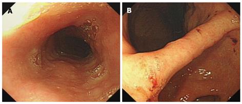
Figure 1 Initial esophagogastroduodenoscopy (EGD) demonstrating normal esophageal mucosa (A) and erosion lesions scattering in the stomach (B).
Figure 2 Second EGD showing submucosal congestion in the esophagus (A), active bleeding in the original biopsy sites (B), and titanium clips used for hemostasis (C).
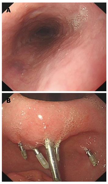
Figure 3 Final EGD showing the absorbed esophageal submucosal congestion (A) and bleeding in the stomach with titanium clips in good condition (B).
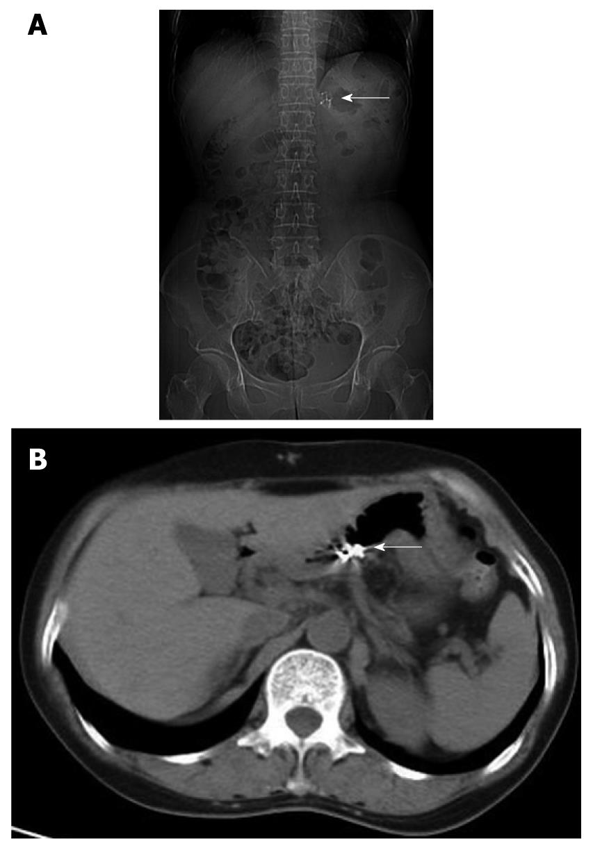
Figure 4 Plain radiography (A) and CT scanning (B) revealing 5 titanium clips in the stomach (arrows).
- Citation: Zhao SL, Li P, Ji M, Zong Y, Zhang ST. Upper gastrointestinal hemorrhage caused by superwarfarin poisoning. World J Gastroenterol 2010; 16(13): 1680-1682
- URL: https://www.wjgnet.com/1007-9327/full/v16/i13/1680.htm
- DOI: https://dx.doi.org/10.3748/wjg.v16.i13.1680


