Copyright
©2010 Baishideng.
World J Gastroenterol. Mar 28, 2010; 16(12): 1537-1540
Published online Mar 28, 2010. doi: 10.3748/wjg.v16.i12.1537
Published online Mar 28, 2010. doi: 10.3748/wjg.v16.i12.1537
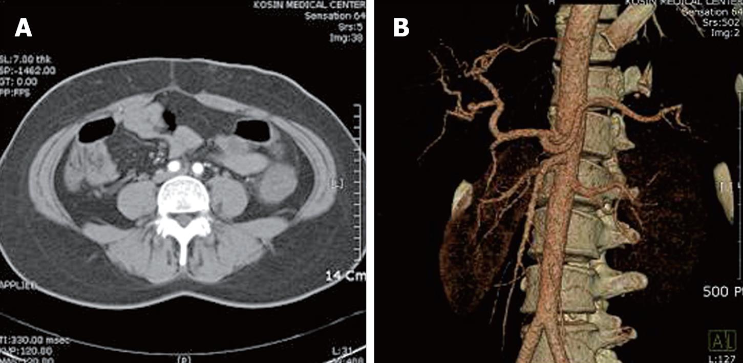
Figure 1 Abdominal computed tomography and angiography.
A: Computed tomography showed diffuse bowel wall edema in the descending colon; B: Angiography showed no evidence of obstruction or stenosis in mesenteric vessels.
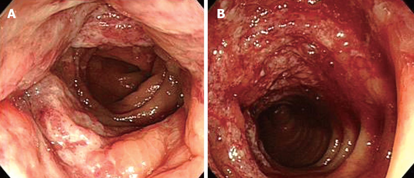
Figure 2 Sigmoidoscopic findings.
Diffuse mucosal edema, erythema (A), friability and submucosal hemorrhage (B) from the splenic flexure to the sigmoid colon are shown.
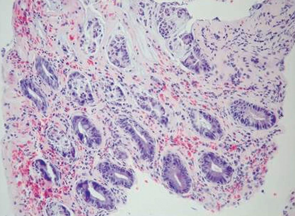
Figure 3 Microscopic findings of colonoscopic biopsy.
Sections showed portions of colonic mucosa revealing sloughing of the surface epithelium and infiltration of mononuclear inflammatory cells in hyalinized lamina propria. (HE, × 200).
- Citation: Kim JB, Moon W, Park SJ, Park MI, Kim KJ, Lee JN, Kang SJ, Jang LL, Chang HK. Ischemic colitis after mesotherapy combined with anti-obesity medications. World J Gastroenterol 2010; 16(12): 1537-1540
- URL: https://www.wjgnet.com/1007-9327/full/v16/i12/1537.htm
- DOI: https://dx.doi.org/10.3748/wjg.v16.i12.1537


