Copyright
©2010 Baishideng.
World J Gastroenterol. Mar 14, 2010; 16(10): 1239-1244
Published online Mar 14, 2010. doi: 10.3748/wjg.v16.i10.1239
Published online Mar 14, 2010. doi: 10.3748/wjg.v16.i10.1239
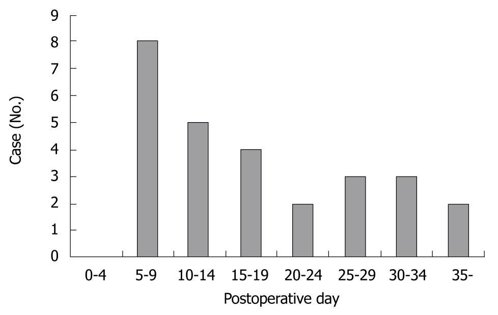
Figure 1 Number of cases based on the onset of pseudoaneurysm rupture.
Figure 2 Flow chart summarizing the outcome of patients with hemorrhage from ruptured pseudoaneurysms.
DIC: Disseminated intravascular coagulation; ARDS: Acute respiratory distress syndrome.
Figure 3 Angiograph.
A: A pseudoaneurysm (arrow) of the gastroduodenal artery; B: A pseudoaneurysm (arrow) of the right hepatic artery; C: A ruptured pseudoaneurysm (arrow) of the right gastric artery.
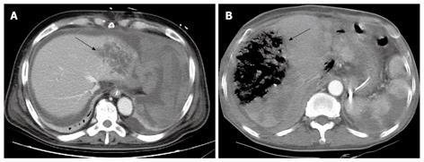
Figure 4 Abdominal CT scan.
A: Liver abscess that may have developed after common hepatic artery embolization (arrow); B: An area of liver infarction following common hepatic artery embolization (arrow).
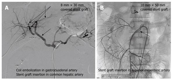
Figure 5 Angiograph.
A: A stent graft of the common hepatic artery placed to manage the gastroduodenal artery pseudoaneurysm (arrow); B: A stent graft insertion for a pseudoaneurysm in the superior mesenteric artery (arrow).
- Citation: Lee HG, Heo JS, Choi SH, Choi DW. Management of bleeding from pseudoaneurysms following pancreaticoduodenectomy. World J Gastroenterol 2010; 16(10): 1239-1244
- URL: https://www.wjgnet.com/1007-9327/full/v16/i10/1239.htm
- DOI: https://dx.doi.org/10.3748/wjg.v16.i10.1239


