Copyright
©2010 Baishideng.
World J Gastroenterol. Jan 7, 2010; 16(1): 76-82
Published online Jan 7, 2010. doi: 10.3748/wjg.v16.i1.76
Published online Jan 7, 2010. doi: 10.3748/wjg.v16.i1.76
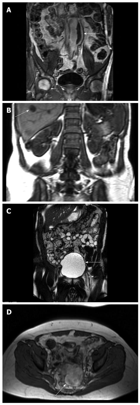
Figure 1 Incidental findings at MRI-enterography.
A: Abdominal aortic aneurysm (arrow). CT scan confirmed the aneurysm and ruled out rupture; B: Atypical hepatic hemangioma (arrow). The results of ultrasound-guided biopsy were benign; C: Large bladder leading to diagnostic work-up and diagnosis of prostate cancer (arrow); D: A lesion with a diameter of 6 cm in the small pelvis (arrow) was confirmed with transvaginal ultrasound. Surgery showed a torquated leiomyoma in the top of the uterus.
- Citation: Jensen MD, Nathan T, Kjeldsen J, Rafaelsen SR. Incidental findings at MRI-enterography in patients with suspected or known Crohn’s disease. World J Gastroenterol 2010; 16(1): 76-82
- URL: https://www.wjgnet.com/1007-9327/full/v16/i1/76.htm
- DOI: https://dx.doi.org/10.3748/wjg.v16.i1.76


