Copyright
©2009 The WJG Press and Baishideng.
World J Gastroenterol. Mar 7, 2009; 15(9): 1141-1143
Published online Mar 7, 2009. doi: 10.3748/wjg.15.1141
Published online Mar 7, 2009. doi: 10.3748/wjg.15.1141
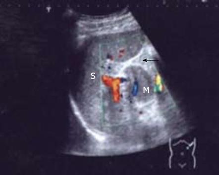
Figure 1 Transverse grayscale sonography image showing a homogeneous and isoechoic nodule (M) in pancreatic tail (arrow) near the spleen hilus with its echogenicity similar to that of the spleen (S).
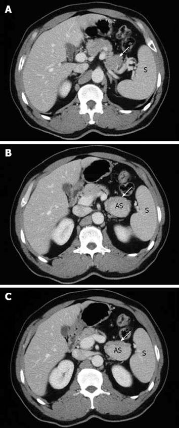
Figure 2 Axial CT image obtained in the portal venous phase showing an ovoid, well-enhanced nodule (AS) in pancreatic tail (arrow).
The density of this lesion was higher than that of the pancreatic parenchyma and similar to that of the spleen (S). However, this finding is similar to that of pancreatic hypervascular tumors.
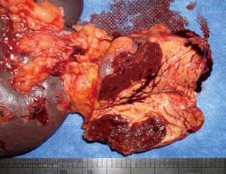
Figure 3 Excision of pancreatic tail and spleen with a well-defined lesion found in pancreatic tail.
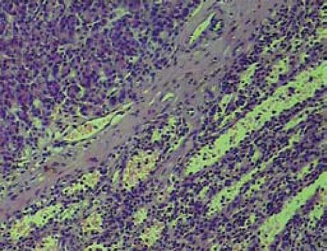
Figure 4 An intrapancreatic accessory spleen confirmed by histological imaging (HE, × 100).
- Citation: Guo W, Han W, Liu J, Jin L, Li JS, Zhang ZT, Wang Y. Intrapancreatic accessory spleen: A case report and review of the literature. World J Gastroenterol 2009; 15(9): 1141-1143
- URL: https://www.wjgnet.com/1007-9327/full/v15/i9/1141.htm
- DOI: https://dx.doi.org/10.3748/wjg.15.1141


