Copyright
©2009 The WJG Press and Baishideng.
World J Gastroenterol. Feb 21, 2009; 15(7): 836-844
Published online Feb 21, 2009. doi: 10.3748/wjg.15.836
Published online Feb 21, 2009. doi: 10.3748/wjg.15.836
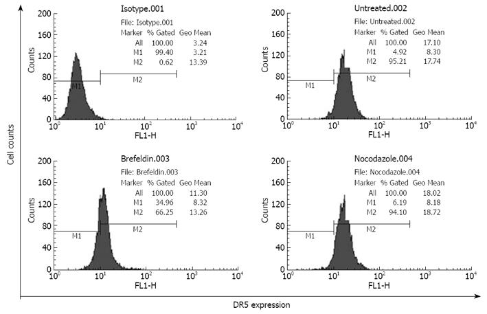
Figure 1 Analysis of DR5 extracellular expression in EC9706 cells by flow cytometry.
Isotype.001: Isotype control; Untreated.002: Extracellular expression of DR5 in the cells pretreated without brefeldin A; Brefeldin.003: Extracellular expression of DR5 in the cells pretreated with brefeldin A; Nocodazole.004: Extracellular expression of DR5 in the cells pretreated with nocodazole for 8 h and without brefeldin A.
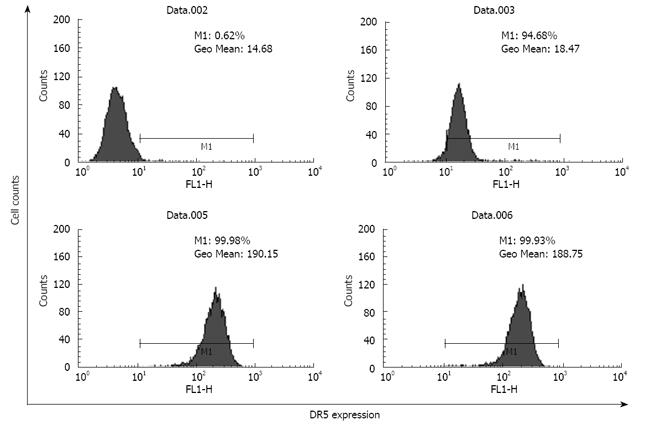
Figure 2 Analysis of total DR5 and extracellular expression in EC9706 cells by flow cytometry.
Data.002: Isotype control; Data.003: Extracellular expression of DR5 in the cells pretreated without brefeldin A; Data.005: Total DR5 expression in the cells pretreated without brefeldin A; Data.006: Total DR5 expression in the cells pretreated with brefeldin A for 30 min before trypsinization.
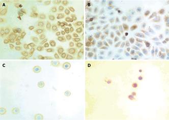
Figure 3 Immunostaining showing expression and localization of DR5 in EC9706 cells.
A: Adherent cells pretreated without brefeldin A; B: Adherent cells pretreated with brefeldin A for 30 min; C: Detached cells not pretreated with brefeldin A before trypsinization; D: Detached cells pretreated with brefeldin A for 30 min before trypsinization (× 400).
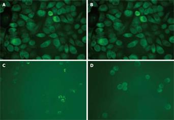
Figure 4 Immunofluorescence showing expression and distribution of DR5 in EC9706 cells.
A: Adherent cells treated without brefeldin A; B: Adherent cells treated with brefeldin A before trypsinization; C: Detached cells not treated with brefeldin A; D: Detached cells treated with brefeldin A for 30 min before trypsinization (× 400).
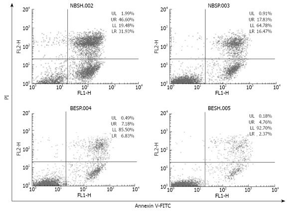
Figure 5 Flow cytomery showing apoptosis of EC9706 cells induced by anti-DR5 agonistic antibody.
NBSH: Detached cells incubated with 2 mg/L mDRA-6 antibody for 12 h; NBSP: Spreading cells incubated with 2 mg/L mDRA-6 antibody for 12 h; BESH: Detached cells incubated in the medium without mDRA-6 antibody for 12 h; BESP: Spreading cells incubated in the medium without mDRA-6 antibody for 12 h.
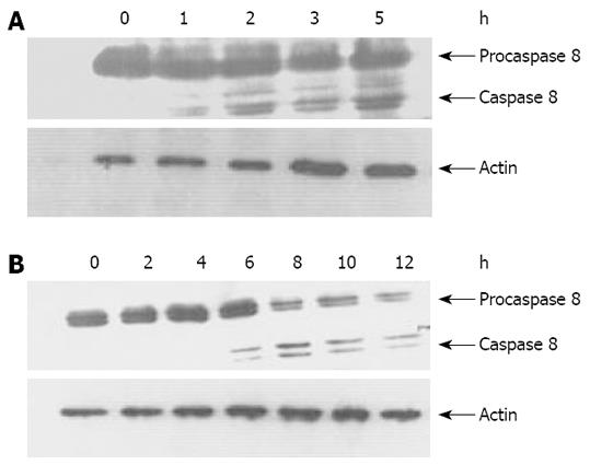
Figure 6 Activation of caspase 8 in EC9706 cells.
A: After trypsinization, temporally detached EC9706 cells were incubated with agonist antibody mDRA-6 for an indicated time at 37°C; B: Spreading EC9706 cells were incubated with agonist antibody mDRA-6 for an indicated time at 37°C. Cell lysates were prepared and Western blotting was performed to analyze caspase 8 activation. The position of procaspase 8 and the active subunits were indicated. Western bloting for actin was utilized as the control of an equal sample loading.
- Citation: Liu GC, Zhang J, Liu SG, Gao R, Long ZF, Tao K, Ma YF. Detachment of esophageal carcinoma cells from extracellular matrix causes relocalization of death receptor 5 and apoptosis. World J Gastroenterol 2009; 15(7): 836-844
- URL: https://www.wjgnet.com/1007-9327/full/v15/i7/836.htm
- DOI: https://dx.doi.org/10.3748/wjg.15.836


