Copyright
©2009 The WJG Press and Baishideng.
World J Gastroenterol. Feb 7, 2009; 15(5): 628-632
Published online Feb 7, 2009. doi: 10.3748/wjg.15.628
Published online Feb 7, 2009. doi: 10.3748/wjg.15.628
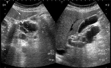
Figure 1 Abdominal ultrosonograpy showing multiple cystic tumors over the pancreatic head area with P-duct dilatation.
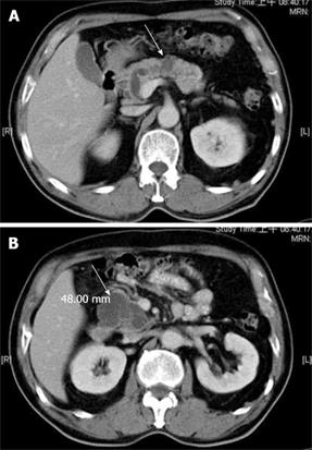
Figure 2 Abdominal computed tomography showing pancreatic head and body cystic tumors (arrow) with no evidence of regional lymphadenopathy or distant metastases.
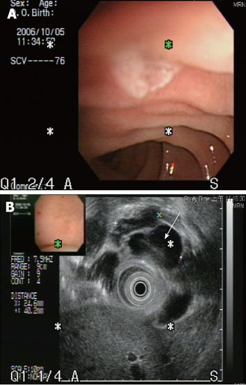
Figure 3 The endoscopic ultrasonogaphy showed that the papilla of Vater was grossly normal without mucin discharge.
However, a cystic tumor (arrow) measuring 4.0 cm x 4.2 cm in size was seen over the pancreatic head. The lining of cysts showed no definite projectile mass and the pancreatic duct was dilated about 0.6 cm in the head and 0.4 cm in the body and tail area.
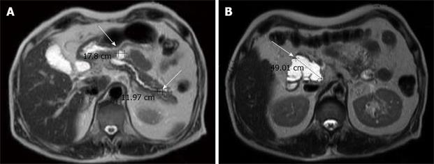
Figure 4 Magnetic resonance imaging showing three cystic lesions in the pancreatic head (4.
9 cm), body (1.8 cm) and tail (1.2 cm), with P-duct dilatation. Mild common bile duct and common hepatic duct dilatation without definite tumor was observed in the liver upon MRI.
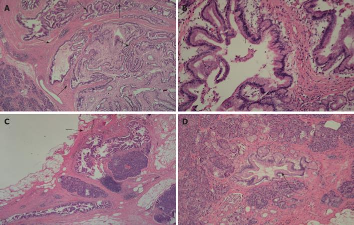
Figure 5 Histopathologic findings (HE × 200).
A: Histopathological examination showed the presence of cystically dilated ducts with formation of papillary growth lined by mucin-secreting columnar epithelium at the level of the pancreatic head; B: The mucin-secreting epithelium reveals stratification of atypical nuclei. No obvious stromal invasion was seen and the features suggested a diagnosis of intraductal papillary mucinous carcinoma, noninvasive type; C: The section of the pancreatic body showed that it was focally involved by the lesion (arrow); D: The section of the pancreatic tail showed the focal presence of atypical ducts.
- Citation: Chiang KC, Hsu JT, Chen HY, Jwo SC, Hwang TL, Jan YY, Yeh CN. Multifocal intraductal papillary mucinous neoplasm of the pancreas-A case report. World J Gastroenterol 2009; 15(5): 628-632
- URL: https://www.wjgnet.com/1007-9327/full/v15/i5/628.htm
- DOI: https://dx.doi.org/10.3748/wjg.15.628


