Copyright
©2009 The WJG Press and Baishideng.
World J Gastroenterol. Dec 7, 2009; 15(45): 5740-5745
Published online Dec 7, 2009. doi: 10.3748/wjg.15.5740
Published online Dec 7, 2009. doi: 10.3748/wjg.15.5740
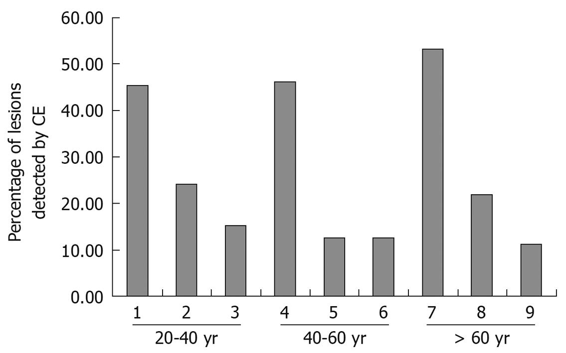
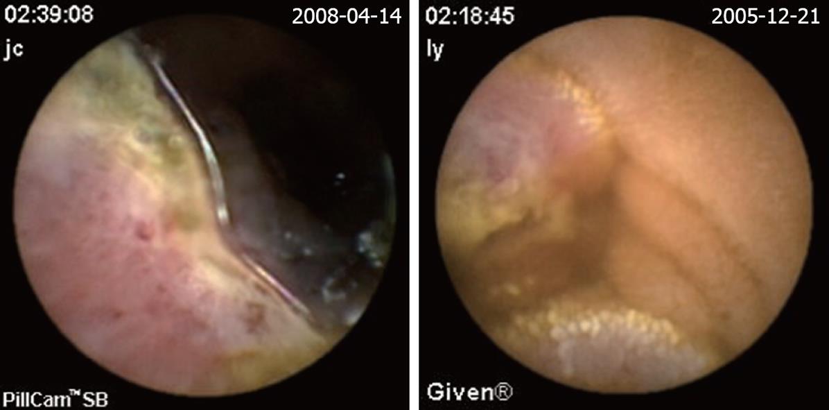
Figure 1 Lesions detected by capsule endoscopy (CE) for different age ranges.
1: Crohn’s disease; 2: SB tumor; 3: Non-specific enteritis; 4: SB tumor; 5: Crohn’s disease; 6: Angioectasias; 7: Angioectasias; 8: SB tumor; 9: Ucler.
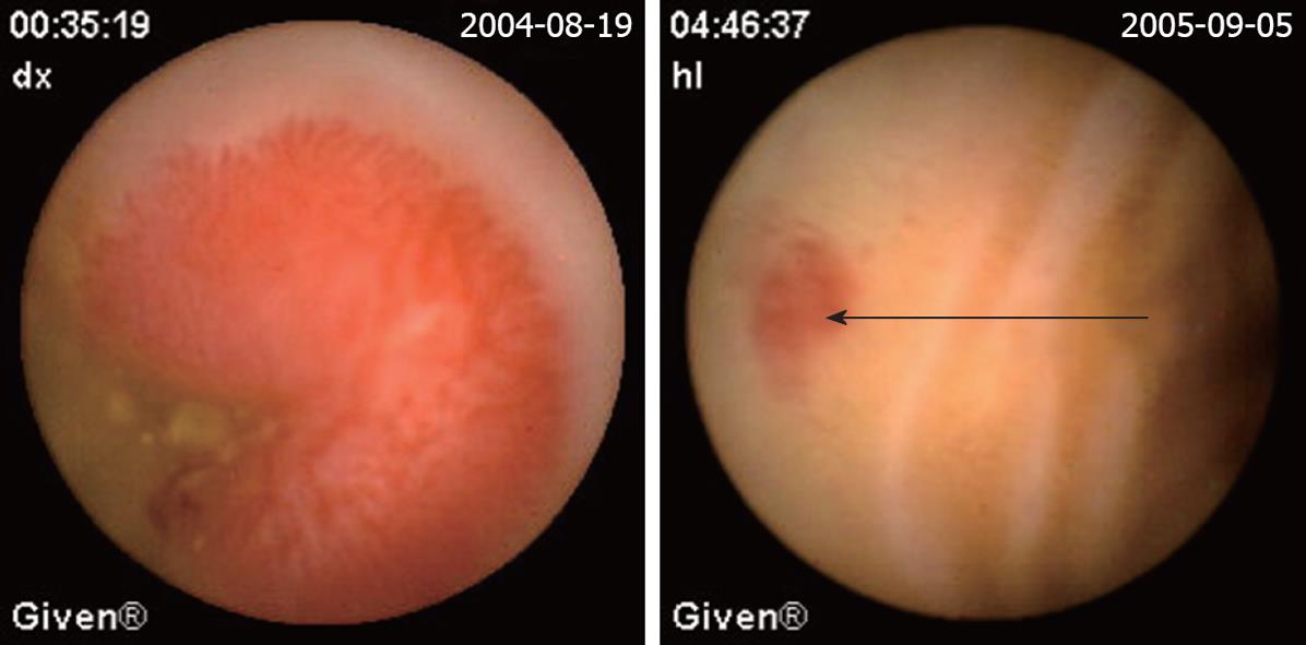
Figure 2 Angioectasias.
The red sopt is the site of angioectasias (arrow).
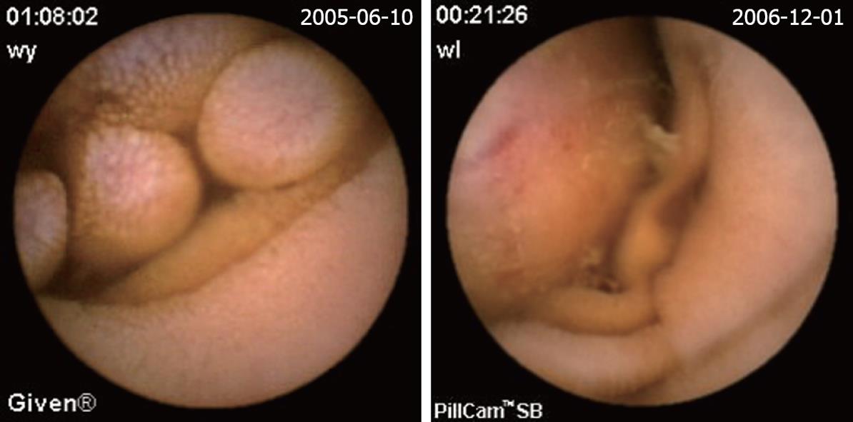
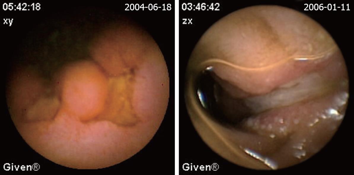
Figure 3 Mesenchymoma.
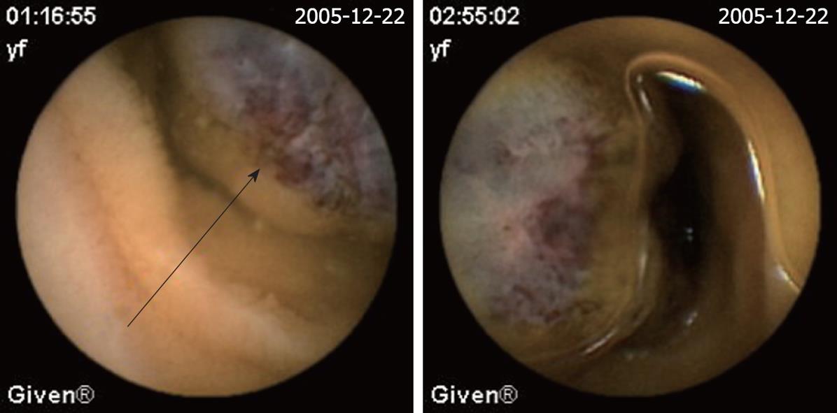
Figure 4 Hemangioma.
The red-blue bubble is the site of hemangioma (arrow).
Figure 5 Lymphoma.
Figure 6 Crohn’s disease.
- Citation: Zhang BL, Fang YH, Chen CX, Li YM, Xiang Z. Single-center experience of 309 consecutive patients with obscure gastrointestinal bleeding. World J Gastroenterol 2009; 15(45): 5740-5745
- URL: https://www.wjgnet.com/1007-9327/full/v15/i45/5740.htm
- DOI: https://dx.doi.org/10.3748/wjg.15.5740


