Copyright
©2009 The WJG Press and Baishideng.
World J Gastroenterol. Sep 28, 2009; 15(36): 4596-4600
Published online Sep 28, 2009. doi: 10.3748/wjg.15.4596
Published online Sep 28, 2009. doi: 10.3748/wjg.15.4596
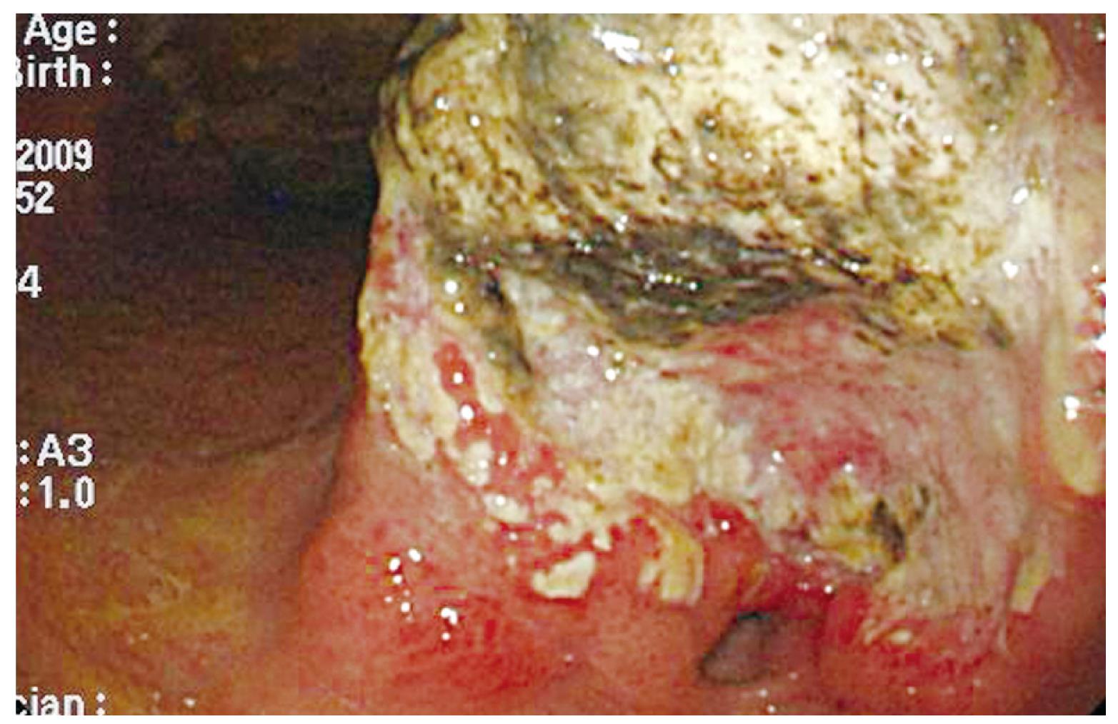
Figure 1 The esophagogastroduodenoscopy (EGD) showed an ulcerated, annular lesion over the gastric antrum.
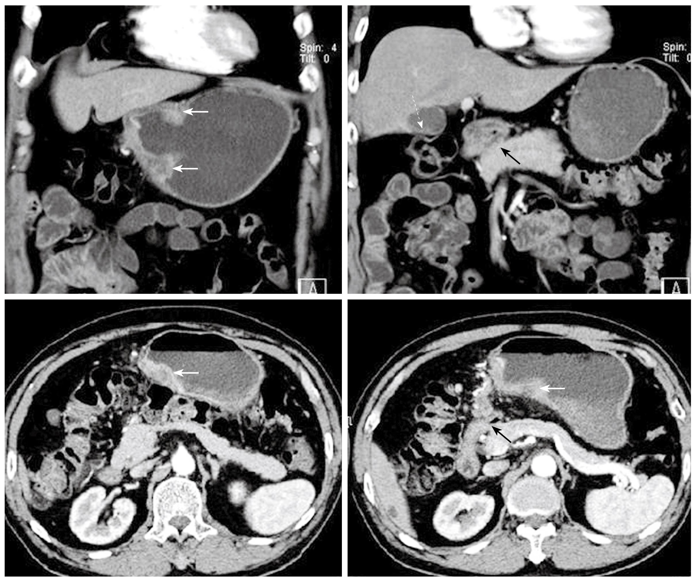
Figure 2 Preoperative abdominal CT images in coronary and transverse sections.
The white solid arrows indicate diffuse wall thickening at the gastric antrum. The white dotted arrow indicates a contracted gallbladder with eccentric wall thickening. The black solid arrows point to the apparent deformity of the duodenal bulb with adhesion to the head of the pancreas. There is no evidence of intra-abdominal metastasis in this study.
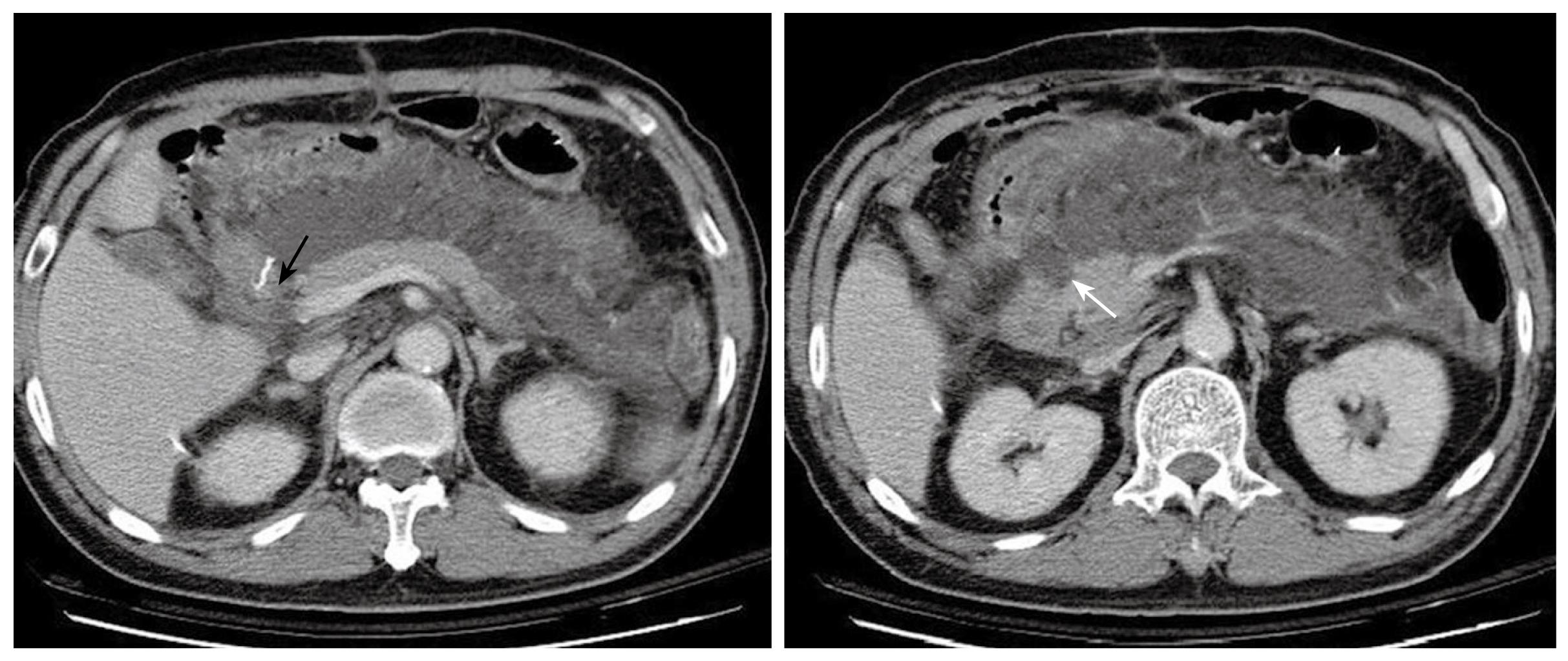
Figure 3 Abdominal CT 8 d after the first operation demonstrates massive fluid accumulation in the peripancreatic area and the lesser sac.
The homogenous fluid extends to the retroperitoneal space. The status of the duodenal stump (black arrow) cannot be clearly assessed. The pancreas is well enhanced and enlarged, and the head shows an uneven and infiltrative margin (white arrow).
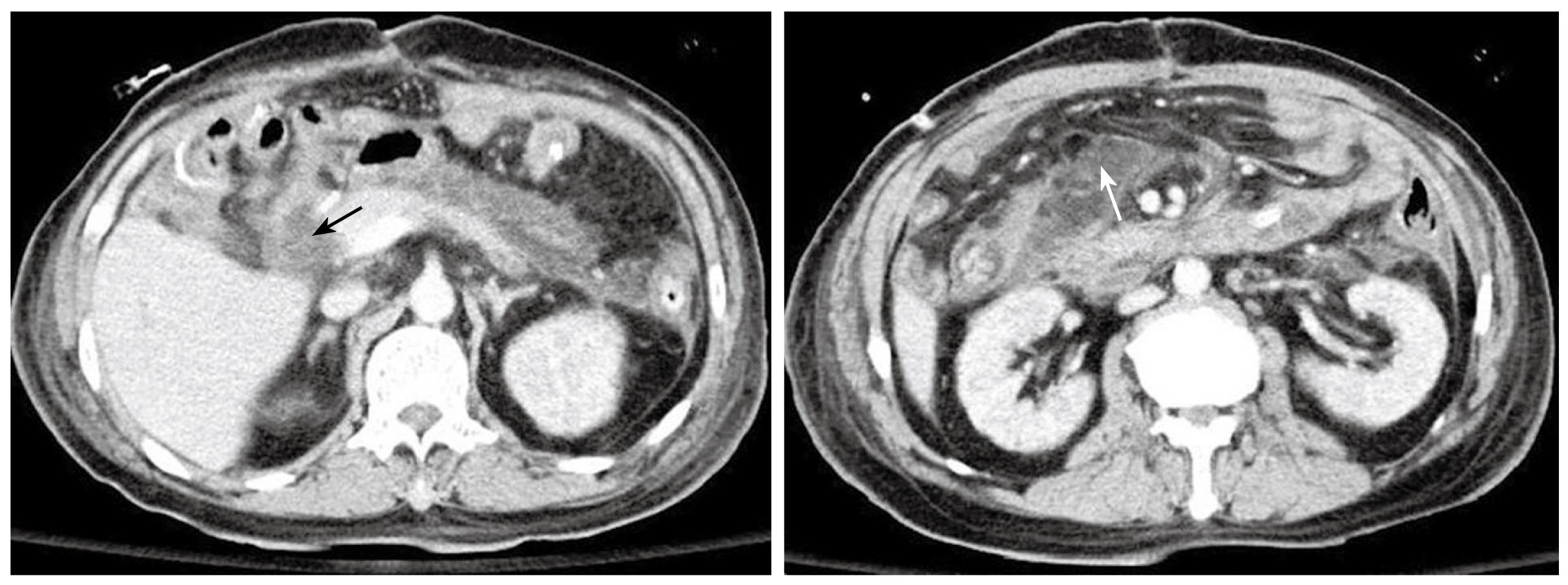
Figure 4 Follow-up abdominal CT after the second operation.
Severe fat stranding is seen. An edematous duodenal stump is observed without a clear fat plane surrounding it. This is highly suspicious of a stump leak (black arrow). The loculated fluid with heterogeneous changes indicates the possibility of an abscess formation (white arrow).
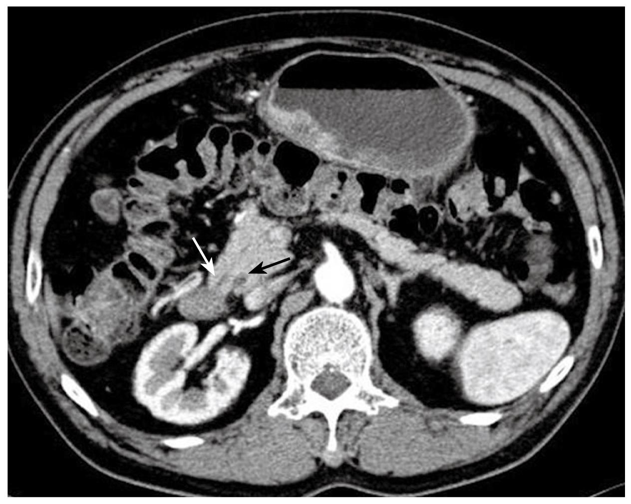
Figure 5 The preoperative CT scan.
The dominant dorsal duct (duct of Santorini) drains into the minor papilla (white arrow). The CBD is seen in the section (black arrow) but the ventral duct (duct of Wirsung) can not be traced. These features are consistent with pancreas divisum.
- Citation: Kuo IM, Wang F, Liu KH, Jan YY. Post-gastrectomy acute pancreatitis in a patient with gastric carcinoma and pancreas divisum. World J Gastroenterol 2009; 15(36): 4596-4600
- URL: https://www.wjgnet.com/1007-9327/full/v15/i36/4596.htm
- DOI: https://dx.doi.org/10.3748/wjg.15.4596


