Copyright
©2009 The WJG Press and Baishideng.
World J Gastroenterol. Sep 28, 2009; 15(36): 4596-4600
Published online Sep 28, 2009. doi: 10.3748/wjg.15.4596
Published online Sep 28, 2009. doi: 10.3748/wjg.15.4596
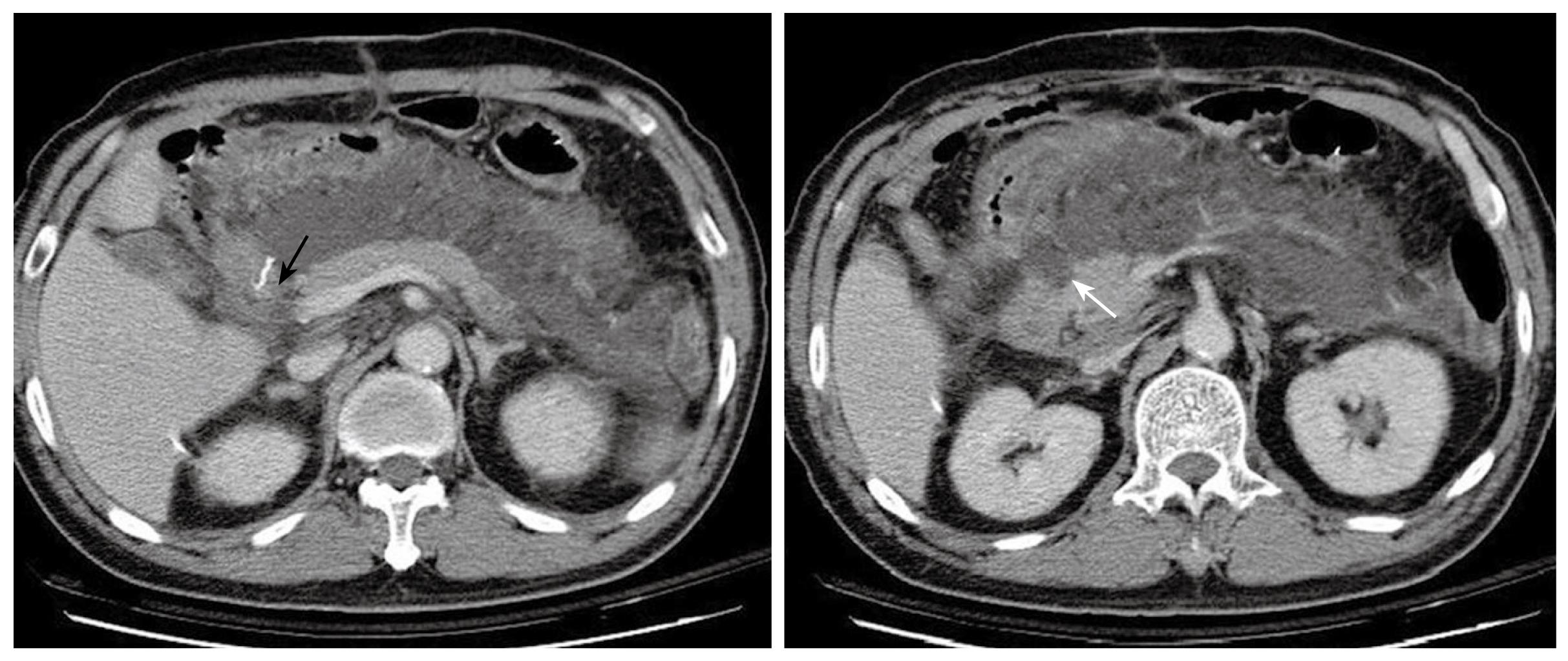
Figure 3 Abdominal CT 8 d after the first operation demonstrates massive fluid accumulation in the peripancreatic area and the lesser sac.
The homogenous fluid extends to the retroperitoneal space. The status of the duodenal stump (black arrow) cannot be clearly assessed. The pancreas is well enhanced and enlarged, and the head shows an uneven and infiltrative margin (white arrow).
- Citation: Kuo IM, Wang F, Liu KH, Jan YY. Post-gastrectomy acute pancreatitis in a patient with gastric carcinoma and pancreas divisum. World J Gastroenterol 2009; 15(36): 4596-4600
- URL: https://www.wjgnet.com/1007-9327/full/v15/i36/4596.htm
- DOI: https://dx.doi.org/10.3748/wjg.15.4596


