Copyright
©2009 The WJG Press and Baishideng.
World J Gastroenterol. Sep 14, 2009; 15(34): 4278-4283
Published online Sep 14, 2009. doi: 10.3748/wjg.15.4278
Published online Sep 14, 2009. doi: 10.3748/wjg.15.4278
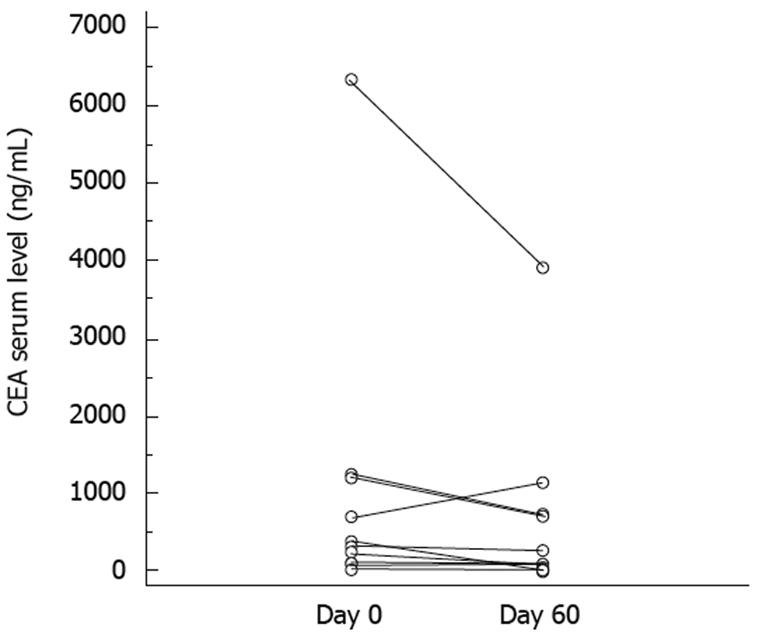
Figure 1 Carcinoembryonic antigen (CEA) serum level before and 2 mo after the beginning of the treatment.
Figure 2 Representative CT scan of patients responding according to Choi criteria.
A: (Patient 5) Contrast-enhanced CT-scan (arterial phase) showed three large liver metastases before inclusion; B: Two months later, a significant decrease in the enhancement of the metastases was observed by CT performed under the same conditions (arterial phase). Although lesions were classified as stable disease using RECIST criteria, they could be considered as a partial response using Choi criteria; C: (Patient 8) Contrast-enhanced CT-scan (arterial phase) showed two latero aortic lymph node metastases. Density was 82.8 UH (arrow). To assure comparability, density was also measured in muscle (72.8 UH) (arrow head); D: After 2 mo, although size of lymph node metastases was stable, there was a significant decrease in density (59 UH) (arrow), which led to a partial response using Choi criteria. In comparison, muscle exhibited no decrease in density (73 UH) (arrow head).
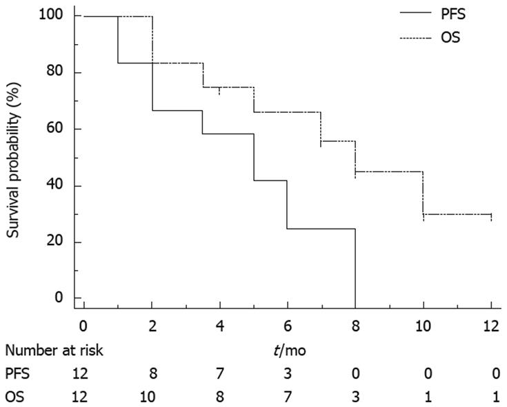
Figure 3 Kaplan–Meier curves of progression-free survival (PFS) and overall survival (OS).
- Citation: Ghiringhelli F, Guiu B, Chauffert B, Ladoire S. Sirolimus, bevacizumab, 5-Fluorouracil and irinotecan for advanced colorectal cancer: A pilot study. World J Gastroenterol 2009; 15(34): 4278-4283
- URL: https://www.wjgnet.com/1007-9327/full/v15/i34/4278.htm
- DOI: https://dx.doi.org/10.3748/wjg.15.4278


