Copyright
©2009 The WJG Press and Baishideng.
World J Gastroenterol. Sep 7, 2009; 15(33): 4193-4195
Published online Sep 7, 2009. doi: 10.3748/wjg.15.4193
Published online Sep 7, 2009. doi: 10.3748/wjg.15.4193
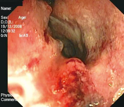
Figure 1 Small cell neuroendocrine carcinoma: endoscopic view.
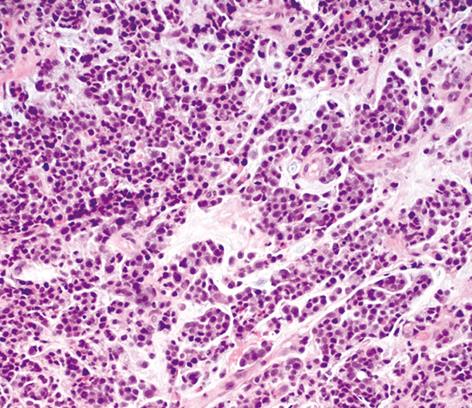
Figure 2 Cells with minimal cytoplasm, fusiform cell shape, finely granular chromatin, small, or absent, nucleoli (HE stain, original magnification × 20).
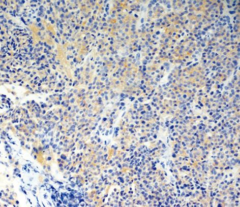
Figure 3 Immunohistochemical analysis reveals neuroendocrine differentiation with positive staining for synaptophysin (original magnification × 20).
- Citation: Grassia R, Bodini P, Dizioli P, Staiano T, Iiritano E, Bianchi G, Buffoli F. Neuroendocrine carcinomas arising in ulcerative colitis: Coincidences or possible correlations? World J Gastroenterol 2009; 15(33): 4193-4195
- URL: https://www.wjgnet.com/1007-9327/full/v15/i33/4193.htm
- DOI: https://dx.doi.org/10.3748/wjg.15.4193


