Copyright
©2009 The WJG Press and Baishideng.
World J Gastroenterol. Aug 21, 2009; 15(31): 3954-3956
Published online Aug 21, 2009. doi: 10.3748/wjg.15.3954
Published online Aug 21, 2009. doi: 10.3748/wjg.15.3954
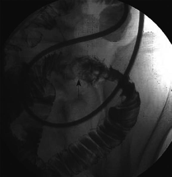
Figure 1 Jejunography using a naso-jejunal tube showed an oval-shaped mass in the jejunum (arrow) and edematous mucosa around the mass.
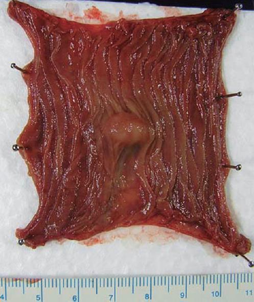
Figure 2 Macroscopic findings of the tumor.
A 14 mm × 11 mm oval-shaped submucosal tumor covered with normal mucosa was observed.
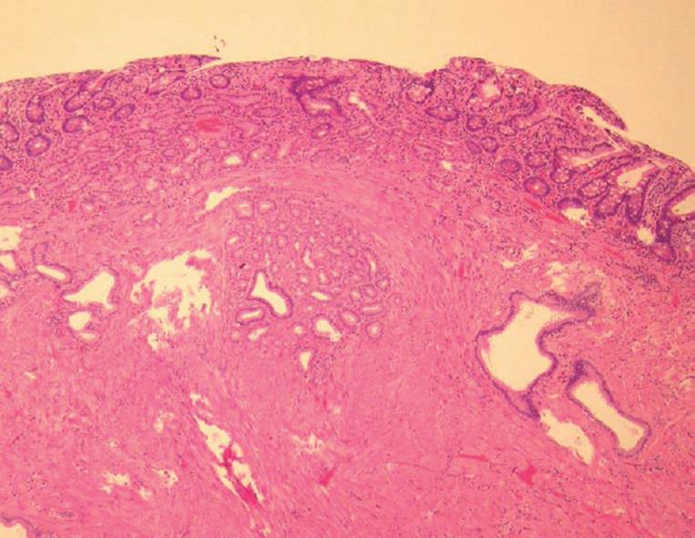
Figure 3 Histological findings of the tumor.
The localized tumor was composed of proliferating ducts and proliferation of smooth muscle bundles without mitotic figures. However, both exocrine acini and endocrine elements were lacking (HE, × 100).
- Citation: Hirasaki S, Kubo M, Inoue A, Miyake Y, Oshiro H. Jejunal small ectopic pancreas developing into jejunojejunal intussusception: A rare cause of ileus. World J Gastroenterol 2009; 15(31): 3954-3956
- URL: https://www.wjgnet.com/1007-9327/full/v15/i31/3954.htm
- DOI: https://dx.doi.org/10.3748/wjg.15.3954


