Copyright
©2009 The WJG Press and Baishideng.
World J Gastroenterol. Aug 14, 2009; 15(30): 3793-3798
Published online Aug 14, 2009. doi: 10.3748/wjg.15.3793
Published online Aug 14, 2009. doi: 10.3748/wjg.15.3793
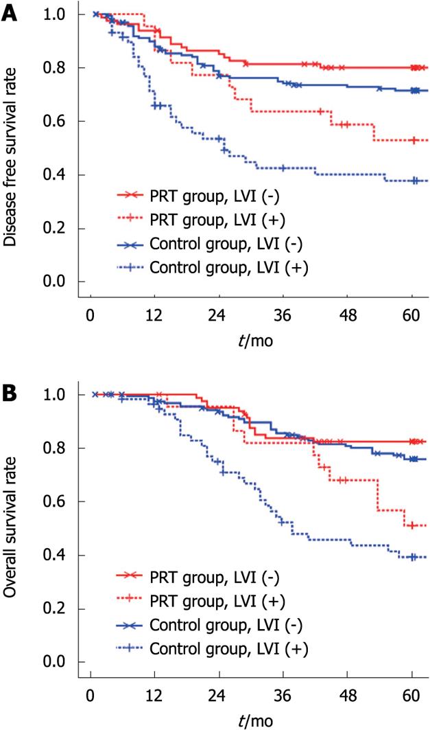
Figure 1 K-M plot of DFS (A) and OS (B) for LVI between the two groups.
The LVI negative patients had a significantly higher 5-year DFS rate (P < 0.05) and OS rate (P < 0.01) than LVI positive patients in both groups (P < 0.05).
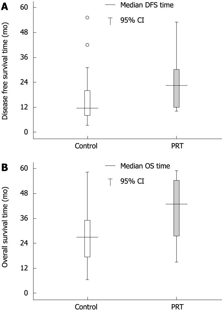
Figure 2 Stem and Leaf plot for comparison of DFS time (A) and OS time (B) for LVI positive patients between the two groups.
Among the 42 (A) and 40 (B) LVI positive patients with disease progression, the patients in the PRT group had a longer median DFS time (P < 0.05).
- Citation: Du CZ, Xue WC, Cai Y, Li M, Gu J. Lymphovascular invasion in rectal cancer following neoadjuvant radiotherapy: A retrospective cohort study. World J Gastroenterol 2009; 15(30): 3793-3798
- URL: https://www.wjgnet.com/1007-9327/full/v15/i30/3793.htm
- DOI: https://dx.doi.org/10.3748/wjg.15.3793


