Copyright
©2009 The WJG Press and Baishideng.
World J Gastroenterol. Aug 7, 2009; 15(29): 3676-3680
Published online Aug 7, 2009. doi: 10.3748/wjg.15.3676
Published online Aug 7, 2009. doi: 10.3748/wjg.15.3676
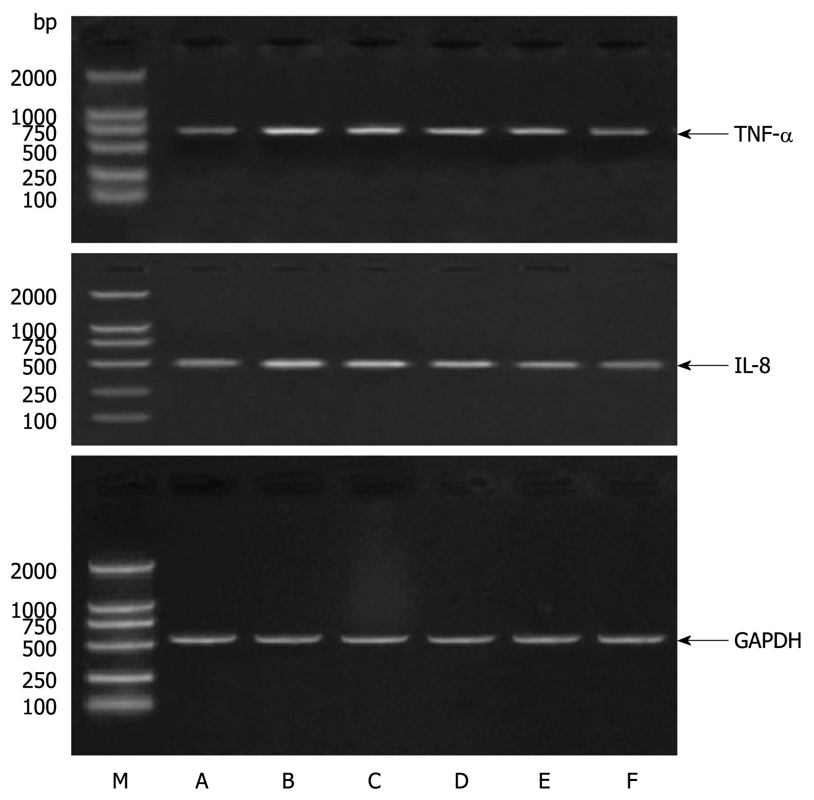
Figure 1 Astragalus mongholicus polysaccharide inhibits TNF-α and IL-8 production in LPS-stimulated rat small intestinal cells.
Intestinal epithelial cells (IEC) were treated with APS for 1 h, and cultured in a medium containing 10 &mgr;g/mL. LPS with APS at different concentrations for 1 h to detect TNF-α and IL-8 mRNAs in the cells by RT-PCR. M: Marker. A: Control group; B: LPS+ 0 &mgr;g/mL APS group; C: LPS+ 50 &mgr;g/mL APS group; D: LPS+ 100 &mgr;g/mL APS group; E: LPS+ 200 &mgr;g/mL APS group; F: LPS+ 500 &mgr;g/mL APS group.
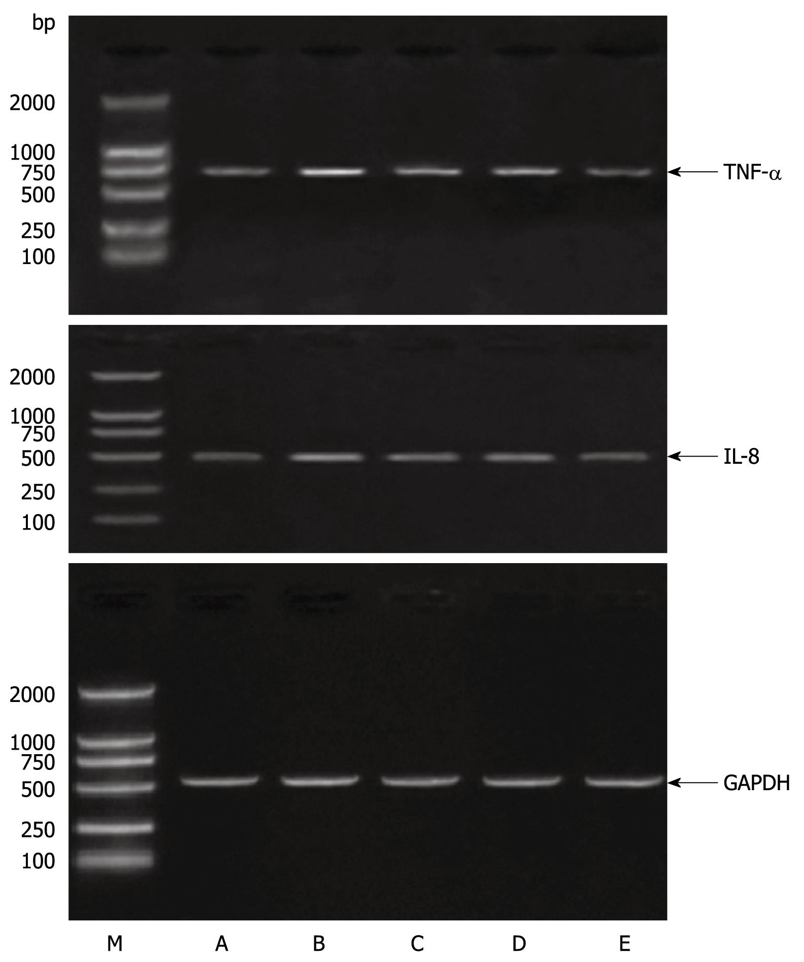
Figure 2 Astragalus mongholicus polysaccharide inhibits TNF-α and IL-8 production in LPS-stimulated rat small intestinal cells.
IEC were treated with APS (500 &mgr;g/mL) for 1 h, and cultured in a medium containing 10 &mgr;g/mL. LPS for up to 4 h to detect TNF-α and IL-8 mRNAs in the cells by RT-PCR. M: Marker. A: Control group; B: LPS+ 0 &mgr;g/mL APS group; C: LPS+ 50 &mgr;g/mL APS group; D: LPS+ 100 &mgr;g/mL APS group; E: LPS+ 200 &mgr;g/mL APS group; F: LPS+ 500 &mgr;g/mL APS group.
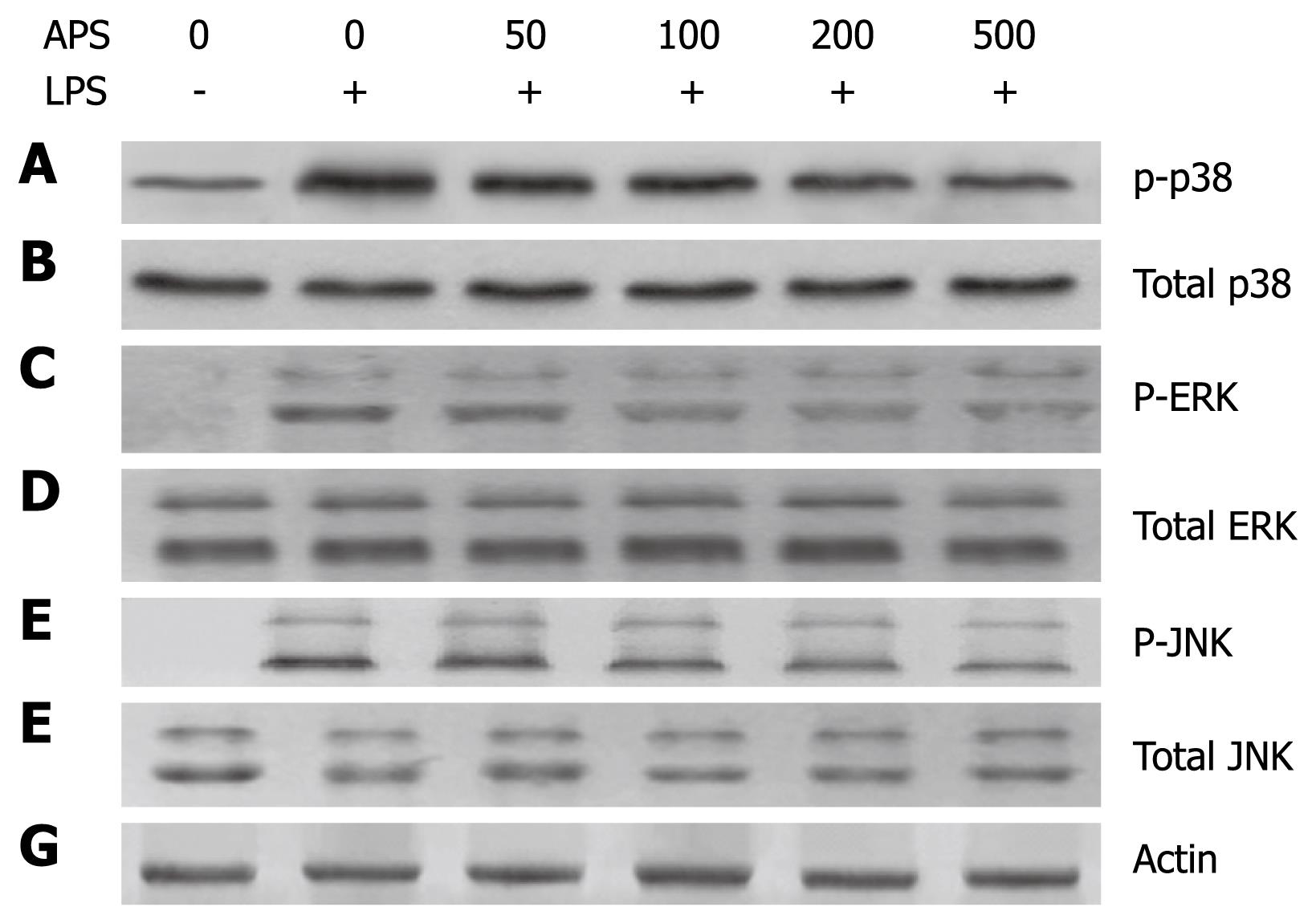
Figure 3 Astragalus mongholicus polysaccharide inhibits p38 phosphorylation but not ERK1/2 or JNK activation in LPS-stimulated rat small intestinal cells.
IEC were treated as described in Figure 1. After incubation in a medium containing 10 &mgr;g/mL LPS with APS at different concentrations of for 1 h, Western blotting analysis was performed to detect phosphorylated p38 (A), total p38 (B), phosphorylated ERK (C), total ERK (D), phosphorylated JNK (E), total JNK (F) and actin (G).
-
Citation: Yuan Y, Sun M, Li KS.
Astragalus mongholicus polysaccharide inhibits lipopolysaccharide-induced production of TNF-α and interleukin-8. World J Gastroenterol 2009; 15(29): 3676-3680 - URL: https://www.wjgnet.com/1007-9327/full/v15/i29/3676.htm
- DOI: https://dx.doi.org/10.3748/wjg.15.3676


