Copyright
©2009 The WJG Press and Baishideng.
World J Gastroenterol. Jul 14, 2009; 15(26): 3309-3311
Published online Jul 14, 2009. doi: 10.3748/wjg.15.3309
Published online Jul 14, 2009. doi: 10.3748/wjg.15.3309
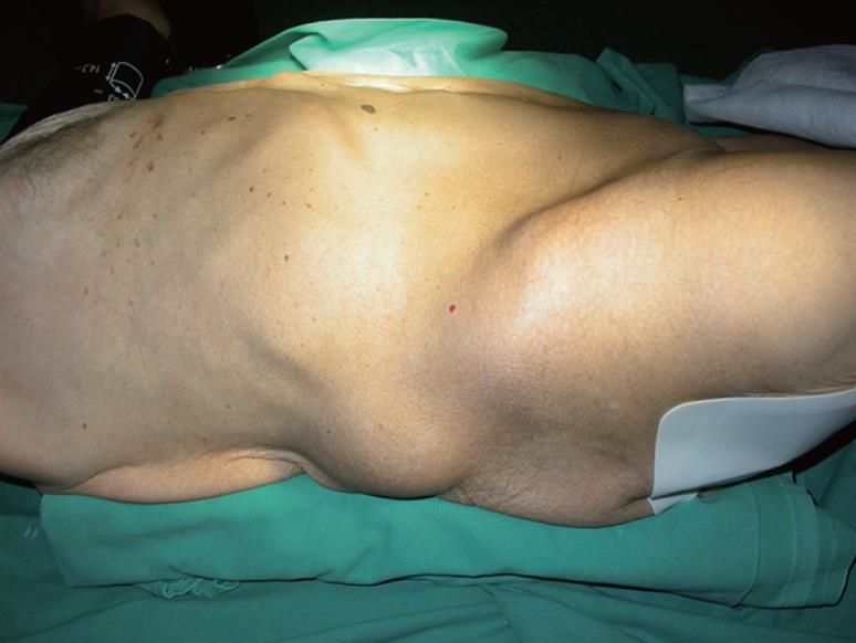
Figure 1 Mass in the right lumbar region.
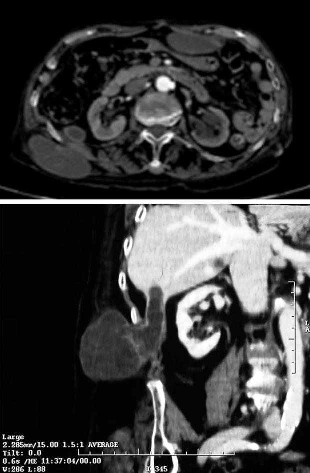
Figure 2 CT.
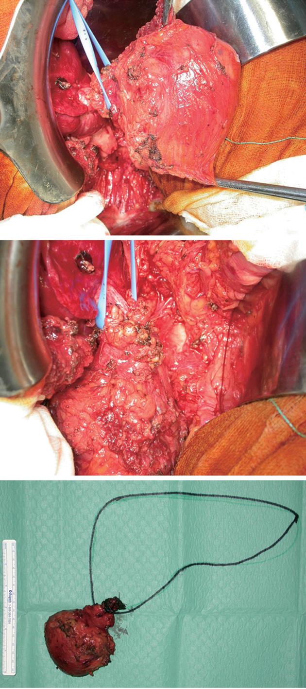
The mass and the peduncle which connected it to the liver, as shown by the scheme with the surgical specimen.
Figure 3 Pedunculated mass arising from the sixth liver segment and growing over the posterior abdominal wall muscles and reaching the subcutaneous tissue.
- Citation: Di Cataldo A, Latino R, Cocuzza A, Li Destri G. Unexplainable development of a hydatid cyst. World J Gastroenterol 2009; 15(26): 3309-3311
- URL: https://www.wjgnet.com/1007-9327/full/v15/i26/3309.htm
- DOI: https://dx.doi.org/10.3748/wjg.15.3309


